8UJV

Crystal structure of human polymerase eta with incoming dCMPnPP nucleotide opposite urea lesion


Experimental Data Snapshot

  • Method: X-RAY DIFFRACTION
  • Resolution: 2.23 Å
  • R-Value Free: 0.222 
  • R-Value Work: 0.166 
  • R-Value Observed: 0.169 

wwPDB Validation   3D Report Full Report


Ligand Structure Quality Assessment 


This is version 1.1 of the entry. See complete history


Literature

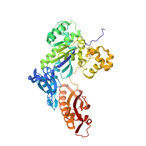
Replication Bypass of the N -(2-Deoxy-d-erythro-pentofuranosyl)-urea DNA Lesion by Human DNA Polymerase eta.

Tomar, R.Li, S.Egli, M.Stone, M.P.

(2024) Biochemistry 63: 754-766

  • DOI: https://doi.org/10.1021/acs.biochem.3c00569
  • Primary Citation of Related Structures:  
    8UJT, 8UJV, 8UJX, 8UK4

  • PubMed Abstract: 

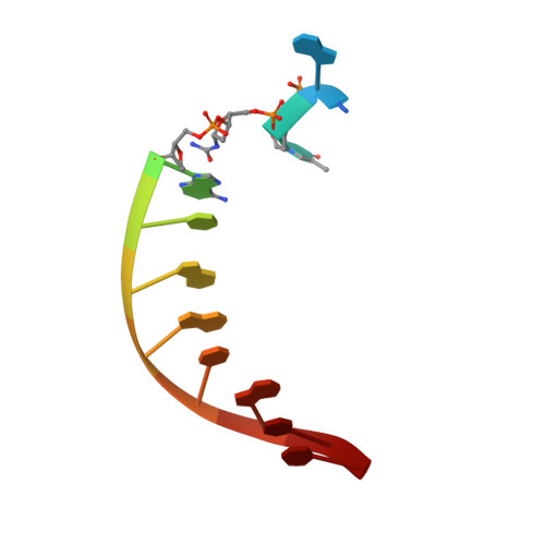
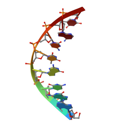
    Urea lesions in DNA arise from thymine glycol (Tg) or 8-oxo-dG; their genotoxicity is thought to arise in part due to their potential to accommodate the insertion of all four dNTPs during error-prone replication. Replication bypass with human DNA polymerase η (hPol η) confirmed that all four dNTPs were inserted opposite urea lesions but with purines exhibiting greater incorporation efficiency. X-ray crystal structures of ternary replication bypass complexes in the presence of Mg 2+ ions with incoming dNTP analogs dAMPnPP, dCMPnPP, dGMPnPP, and dTMPnPP bound opposite urea lesions (hPol η·DNA·dNMPnPP complexes) revealed all were accommodated by hPol η. In each, the Watson-Crick face of the dNMPnPP was paired with the urea lesion, exploiting the ability of the amine and carbonyl groups of the urea to act as H-bond donors or acceptors, respectively. With incoming dAMPnPP or dGMPnPP, the distance between the imino nitrogen of urea and the N9 atoms of incoming dNMPnPP approximated the canonical distance of 9 Å in B-DNA. With incoming dCMPnPP or dTMPnPP, the corresponding distance of about 7 Å was less ideal. Improved base-stacking interactions were also observed with incoming purines vs pyrimidines. Nevertheless, in each instance, the α-phosphate of incoming dNMPnPPs was close to the 3'-hydroxyl group of the primer terminus, consistent with the catalysis of nucleotidyl transfer and the observation that all four nucleotides could be inserted opposite urea lesions. Preferential insertion of purines by hPol η may explain, in part, why the urea-directed spectrum of mutations arising from Tg vs 8-oxo-dG lesions differs.


  • Organizational Affiliation

    Department of Chemistry, Vanderbilt Ingram Cancer Center, and Vanderbilt Center for Structural Biology, Vanderbilt University, Nashville, Tennessee 37235, United States.


Macromolecules

Find similar proteins by:  (by identity cutoff)  |  3D Structure
Entity ID: 1
MoleculeChains Sequence LengthOrganismDetailsImage
DNA polymerase eta435Homo sapiensMutation(s): 0 
Gene Names: POLHRAD30RAD30AXPV
EC: 2.7.7.7
UniProt & NIH Common Fund Data Resources
Find proteins for Q9Y253 (Homo sapiens)
Explore Q9Y253 
Go to UniProtKB:  Q9Y253
PHAROS:  Q9Y253
GTEx:  ENSG00000170734 
Entity Groups  
Sequence Clusters30% Identity50% Identity70% Identity90% Identity95% Identity100% Identity
UniProt GroupQ9Y253
Sequence Annotations
Expand
  • Reference Sequence

Find similar nucleic acids by:  Sequence   |   3D Structure  

Entity ID: 2
MoleculeChains LengthOrganismImage
DNA (5'-D(*CP*AP*TP*(UD8)P*AP*TP*GP*AP*CP*GP*CP*T)-3')B [auth T]12synthetic construct
Sequence Annotations
Expand
  • Reference Sequence

Find similar nucleic acids by:  Sequence   |   3D Structure  

Entity ID: 3
MoleculeChains LengthOrganismImage
DNA (5'-D(*AP*GP*CP*GP*TP*CP*AP*T)-3')C [auth P]8synthetic construct
Sequence Annotations
Expand
  • Reference Sequence
Experimental Data & Validation

Experimental Data

  • Method: X-RAY DIFFRACTION
  • Resolution: 2.23 Å
  • R-Value Free: 0.222 
  • R-Value Work: 0.166 
  • R-Value Observed: 0.169 
  • Space Group: P 61
Unit Cell:
Length ( Å )Angle ( ˚ )
a = 98.951α = 90
b = 98.951β = 90
c = 81.498γ = 120
Software Package:
Software NamePurpose
HKL-2000data reduction
HKL-2000data scaling
Cootmodel building
PHASERphasing
PHENIXrefinement

Structure Validation

View Full Validation Report


Ligand Structure Quality Assessment 


Entry History & Funding Information

Deposition Data


Funding OrganizationLocationGrant Number
National Institutes of Health/National Cancer Institute (NIH/NCI)United StatesR01 ES-029357

Revision History  (Full details and data files)

  • Version 1.0: 2024-03-20
    Type: Initial release
  • Version 1.1: 2024-04-03
    Changes: Database references