8UJA

T33-fn10 - Designed Tetrahedral Protein Cage Using Fragment-based Hydrogen Bond Networks


Experimental Data Snapshot

  • Method: X-RAY DIFFRACTION
  • Resolution: 6.00 Å
  • R-Value Free: 0.248 
  • R-Value Work: 0.214 
  • R-Value Observed: 0.218 

wwPDB Validation   3D Report Full Report


This is version 1.1 of the entry. See complete history


Literature

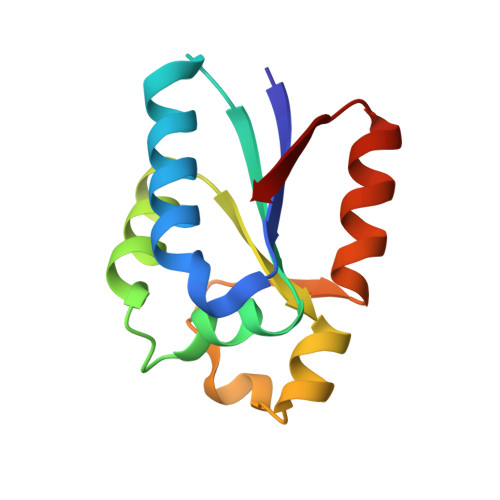
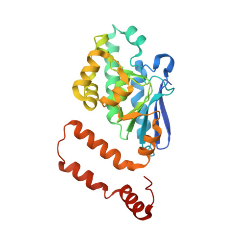
A suite of designed protein cages using machine learning and protein fragment-based protocols.

Meador, K.Castells-Graells, R.Aguirre, R.Sawaya, M.R.Arbing, M.A.Sherman, T.Senarathne, C.Yeates, T.O.

(2024) Structure 

  • DOI: https://doi.org/10.1016/j.str.2024.02.017
  • Primary Citation of Related Structures:  
    8UF0, 8UI2, 8UJA, 8UKM, 8UMP, 8UMR, 8UN1

  • PubMed Abstract: 

    Designed protein cages and related materials provide unique opportunities for applications in biotechnology and medicine, but their creation remains challenging. Here, we apply computational approaches to design a suite of tetrahedrally symmetric, self-assembling protein cages. For the generation of docked conformations, we emphasize a protein fragment-based approach, while for sequence design of the de novo interface, a comparison of knowledge-based and machine learning protocols highlights the power and increased experimental success achieved using ProteinMPNN. An analysis of design outcomes provides insights for improving interface design protocols, including prioritizing fragment-based motifs, balancing interface hydrophobicity and polarity, and identifying preferred polar contact patterns. In all, we report five structures for seven protein cages, along with two structures of intermediate assemblies, with the highest resolution reaching 2.0 Å using cryo-EM. This set of designed cages adds substantially to the body of available protein nanoparticles, and to methodologies for their creation.


  • Organizational Affiliation

    Department of Chemistry and Biochemistry, University of California, Los Angeles, CA 90095, USA.


Macromolecules
Find similar proteins by:  (by identity cutoff)  |  3D Structure
Entity ID: 1
MoleculeChains Sequence LengthOrganismDetailsImage
T33-fn10: engineered DrsE like sulfur reductase
A, C, E, G, I
A, C, E, G, I, K, M, O
108Sulfurisphaera tokodaii str. 7Mutation(s): 0 
Entity Groups  
Sequence Clusters30% Identity50% Identity70% Identity90% Identity95% Identity100% Identity
Sequence Annotations
Expand
  • Reference Sequence
Find similar proteins by:  (by identity cutoff)  |  3D Structure
Entity ID: 2
MoleculeChains Sequence LengthOrganismDetailsImage
T33-fn10: engineered enoyl-CoA hydratase/isomerase
B, D, F, H, J
B, D, F, H, J, L, N, P
258Novosphingobium aromaticivorans DSM 12444Mutation(s): 6 
Gene Names: Saro_3457
UniProt
Find proteins for A4XEF6 (Novosphingobium aromaticivorans (strain ATCC 700278 / DSM 12444 / CCUG 56034 / CIP 105152 / NBRC 16084 / F199))
Explore A4XEF6 
Go to UniProtKB:  A4XEF6
Entity Groups  
Sequence Clusters30% Identity50% Identity70% Identity90% Identity95% Identity100% Identity
UniProt GroupA4XEF6
Sequence Annotations
Expand
  • Reference Sequence
Experimental Data & Validation

Experimental Data

  • Method: X-RAY DIFFRACTION
  • Resolution: 6.00 Å
  • R-Value Free: 0.248 
  • R-Value Work: 0.214 
  • R-Value Observed: 0.218 
  • Space Group: P 21 3
Unit Cell:
Length ( Å )Angle ( ˚ )
a = 225.63α = 90
b = 225.63β = 90
c = 225.63γ = 90
Software Package:
Software NamePurpose
PHENIXrefinement
XDSdata reduction
XDSdata scaling
PHASERphasing

Structure Validation

View Full Validation Report


Entry History & Funding Information

Deposition Data


Funding OrganizationLocationGrant Number
National Institutes of Health/National Institute of General Medical Sciences (NIH/NIGMS)United StatesR01GM129854

Revision History  (Full details and data files)

  • Version 1.0: 2024-03-06
    Type: Initial release
  • Version 1.1: 2024-04-03
    Changes: Database references