8U8Y | pdb_00008u8y

Human retinal variant phosphomimetic IMPDH1(546)-S477D filament bound by ATP, IMP, and NAD+, interface-centered

  • Classification: OXIDOREDUCTASE
  • Organism(s): Homo sapiens
  • Expression System: Escherichia coli BL21(DE3)
  • Mutation(s): Yes 

  • Deposited: 2023-09-18 Released: 2024-01-31 
  • Deposition Author(s): Calise, S.J., Kollman, J.M.
  • Funding Organization(s): National Institutes of Health/National Institute of General Medical Sciences (NIH/NIGMS), National Institutes of Health/National Eye Institute (NIH/NEI), National Institutes of Health/Office of the Director, Helen Hay Whitney Foundation

Experimental Data Snapshot

  • Method: ELECTRON MICROSCOPY
  • Resolution: 2.10 Å
  • Aggregation State: FILAMENT 
  • Reconstruction Method: SINGLE PARTICLE 

wwPDB Validation 3D Report Full Report

Validation slider image for 8U8Y

This is version 1.1 of the entry. See complete history

Literature

Light-sensitive phosphorylation regulates retinal IMPDH1 activity and filament assembly.

Calise, S.J.O'Neill, A.G.Burrell, A.L.Dickinson, M.S.Molfino, J.Clarke, C.Quispe, J.Sokolov, D.Buey, R.M.Kollman, J.M.

(2024) J Cell Biol 223

  • DOI: https://doi.org/10.1083/jcb.202310139
  • Primary Citation Related Structures: 
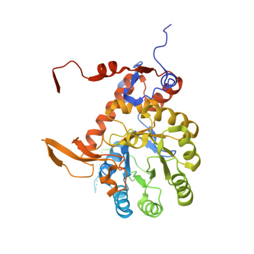
    8U7M, 8U7Q, 8U7V, 8U8O, 8U8Y

  • PubMed Abstract: 

    Inosine monophosphate dehydrogenase (IMPDH) is the rate-limiting enzyme in guanosine triphosphate (GTP) synthesis and assembles into filaments in cells, which desensitizes the enzyme to feedback inhibition and boosts nucleotide production. The vertebrate retina expresses two splice variants IMPDH1(546) and IMPDH1(595). In bovine retinas, residue S477 is preferentially phosphorylated in the dark, but the effects on IMPDH1 activity and regulation are unclear. Here, we generated phosphomimetic mutants to investigate structural and functional consequences of S477 phosphorylation. The S477D mutation resensitized both variants to GTP inhibition but only blocked assembly of IMPDH1(595) filaments. Cryo-EM structures of both variants showed that S477D specifically blocks assembly of a high-activity assembly interface, still allowing assembly of low-activity IMPDH1(546) filaments. Finally, we discovered that S477D exerts a dominant-negative effect in cells, preventing endogenous IMPDH filament assembly. By modulating the structure and higher-order assembly of IMPDH, S477 phosphorylation acts as a mechanism for downregulating retinal GTP synthesis in the dark when nucleotide turnover is decreased.


  • Organizational Affiliation
    • Department of Biochemistry, University of Washington, Seattle, WA, USA.

Macromolecule Content 

  • Total Structure Weight: 476.44 kDa 
  • Atom Count: 22,616 
  • Modeled Residue Count: 2,936 
  • Deposited Residue Count: 4,384 
  • Unique protein chains: 1

Macromolecules

Find similar proteins by:|  3D Structure
Entity ID: 1
MoleculeChains  Sequence LengthOrganismDetailsImage
Inosine-5'-monophosphate dehydrogenase 1
A, B, C, D, E
A, B, C, D, E, F, G, H
548Homo sapiensMutation(s): 38 
Gene Names: IMPDH1IMPD1
EC: 1.1.1.205
UniProt & NIH Common Fund Data Resources
Find proteins for P20839 (Homo sapiens)
Explore P20839 
Go to UniProtKB:  P20839
PHAROS:  P20839
GTEx:  ENSG00000106348 
Entity Groups
Sequence Clusters30% Identity50% Identity70% Identity90% Identity95% Identity100% Identity
UniProt GroupP20839
Sequence Annotations
Expand
Reference Sequence

Small Molecules

Ligands 2 Unique
IDChains Name / Formula / InChI Key2D Diagram3D Interactions
NAD
(Subject of Investigation/LOI)

Query on NAD


Download:Ideal Coordinates CCD File
J [auth A]
L [auth B]
N [auth C]
P [auth D]
R [auth E]
J [auth A],
L [auth B],
N [auth C],
P [auth D],
R [auth E],
T [auth F],
V [auth G],
X [auth H]
NICOTINAMIDE-ADENINE-DINUCLEOTIDE
C21 H27 N7 O14 P2
BAWFJGJZGIEFAR-NNYOXOHSSA-N
IMP
(Subject of Investigation/LOI)

Query on IMP


Download:Ideal Coordinates CCD File
I [auth A]
K [auth B]
M [auth C]
O [auth D]
Q [auth E]
I [auth A],
K [auth B],
M [auth C],
O [auth D],
Q [auth E],
S [auth F],
U [auth G],
W [auth H]
INOSINIC ACID
C10 H13 N4 O8 P
GRSZFWQUAKGDAV-KQYNXXCUSA-N

Experimental Data & Validation

Experimental Data

  • Method: ELECTRON MICROSCOPY
  • Resolution: 2.10 Å
  • Aggregation State: FILAMENT 
  • Reconstruction Method: SINGLE PARTICLE 
EM Software:
TaskSoftware PackageVersion
MODEL REFINEMENTPHENIX1.20.1_4487:
RECONSTRUCTIONcryoSPARC

Structure Validation

View Full Validation Report


Entry History 

& Funding Information

Deposition Data


Funding OrganizationLocationGrant Number
National Institutes of Health/National Institute of General Medical Sciences (NIH/NIGMS)United StatesR01GM118396
National Institutes of Health/National Institute of General Medical Sciences (NIH/NIGMS)United StatesR35GM149542
National Institutes of Health/National Eye Institute (NIH/NEI)United StatesR21EY031546
National Institutes of Health/Office of the DirectorUnited StatesS10OD032290
Helen Hay Whitney FoundationUnited States--

Revision History  (Full details and data files)

  • Version 1.0: 2024-01-31
    Type: Initial release
  • Version 1.1: 2024-02-14
    Changes: Database references