Improving Protein Expression, Stability, and Function with ProteinMPNN.
Sumida, K.H., Nunez-Franco, R., Kalvet, I., Pellock, S.J., Wicky, B.I.M., Milles, L.F., Dauparas, J., Wang, J., Kipnis, Y., Jameson, N., Kang, A., De La Cruz, J., Sankaran, B., Bera, A.K., Jimenez-Oses, G., Baker, D.(2024) J Am Chem Soc 146: 2054-2061
- PubMed: 38194293 Search on PubMedSearch on PubMed Central
- DOI: https://doi.org/10.1021/jacs.3c10941
- Primary Citation Related Structures:
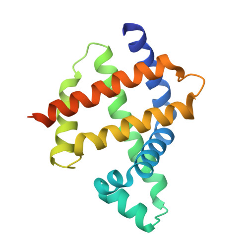
8U5A - PubMed Abstract:
Natural proteins are highly optimized for function but are often difficult to produce at a scale suitable for biotechnological applications due to poor expression in heterologous systems, limited solubility, and sensitivity to temperature. Thus, a general method that improves the physical properties of native proteins while maintaining function could have wide utility for protein-based technologies. Here, we show that the deep neural network ProteinMPNN, together with evolutionary and structural information, provides a route to increasing protein expression, stability, and function. For both myoglobin and tobacco etch virus (TEV) protease, we generated designs with improved expression, elevated melting temperatures, and improved function. For TEV protease, we identified multiple designs with improved catalytic activity as compared to the parent sequence and previously reported TEV variants. Our approach should be broadly useful for improving the expression, stability, and function of biotechnologically important proteins.
- Department of Chemistry, University of Washington, Seattle, Washington 98195, United States.
Organizational Affiliation: