Distal protein-protein interactions contribute to nirmatrelvir resistance.
Lewandowski, E.M., Zhang, X., Tan, H., Jaskolka-Brown, A., Kohaal, N., Frazier, A., Madsen, J.J., Jacobs, L.M.C., Wang, J., Chen, Y.(2025) Nat Commun 16: 1266-1266
- PubMed: 39893201 Search on PubMedSearch on PubMed Central
- DOI: https://doi.org/10.1038/s41467-025-56651-x
- Primary Citation Related Structures:
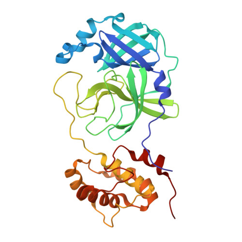
8U25, 8U4Y - PubMed Abstract:
SARS-CoV-2 main protease, M pro , is responsible for processing the viral polyproteins into individual proteins, including the protease itself. M pro is a key target of anti-COVID-19 therapeutics such as nirmatrelvir (the active component of Paxlovid). Resistance mutants identified clinically and in viral passage assays contain a combination of active site mutations (e.g., E166V, E166A, L167F), which reduce inhibitor binding and enzymatic activity, and non-active site mutations (e.g., P252L, T21I, L50F), which restore the fitness of viral replication. To probe the role of the non-active site mutations in fitness rescue, here we use an M pro triple mutant (L50F/E166A/L167F) that confers nirmatrelvir drug resistance with a viral fitness level similar to the wild-type. By comparing peptide and full-length M pro protein as substrates, we demonstrate that the binding of M pro substrate involves more than residues in the active site. Particularly, L50F and other non-active site mutations can enhance the M pro dimer-dimer interactions and help place the nsp5-6 substrate at the enzyme catalytic center. The structural and enzymatic activity data of M pro L50F, L50F/E166A/L167F, and others underscore the importance of considering the whole substrate protein in studying M pro and substrate interactions, and offers important insights into M pro function, resistance development, and inhibitor design.
- Department of Molecular Medicine, Morsani College of Medicine, University of South Florida, Tampa, FL, USA.
Organizational Affiliation: