


De novo Design of Near Infrared Fluorescent Proteins
Xu, C., Liu, Y., Baker, D.To be published.
Experimental Data Snapshot
Starting Model: other
View more details
wwPDB Validation 3D Report Full Report

Entity ID: 1 | |||||
|---|---|---|---|---|---|
| Molecule | Chains | Sequence Length | Organism | Details | Image |
| Designed Near Infrared Fluorescent Protein MC7BP34 | 379 | synthetic construct | Mutation(s): 0 | 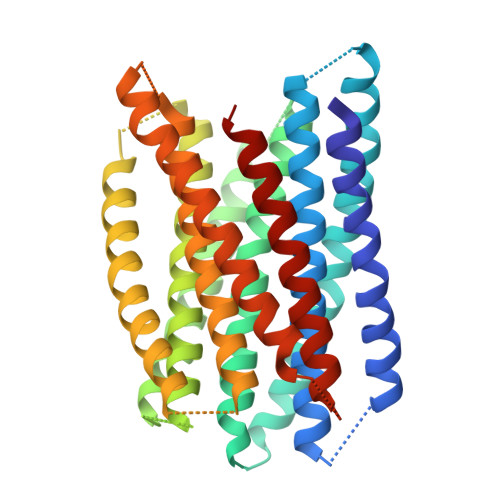 | |
| Length ( Å ) | Angle ( ˚ ) |
|---|---|
| a = 54.192 | α = 90 |
| b = 80.443 | β = 94.261 |
| c = 72.631 | γ = 90 |
| Software Name | Purpose |
|---|---|
| PHENIX | refinement |
| iMOSFLM | data reduction |
| Aimless | data scaling |
| PHASER | phasing |
| Funding Organization | Location | Grant Number |
|---|---|---|
| Howard Hughes Medical Institute (HHMI) | United States | -- |


