8U06 | pdb_00008u06

Imine reductase RedE bound with NADP+ and arcyriaflavin A (primary site)


Experimental Data Snapshot

  • Method: X-RAY DIFFRACTION
  • Resolution: 1.60 Å
  • R-Value Free: 
    0.189 (Depositor), 0.190 (DCC) 
  • R-Value Work: 
    0.155 (Depositor), 0.155 (DCC) 
  • R-Value Observed: 
    0.156 (Depositor) 

Starting Model: experimental
View more details

wwPDB Validation 3D Report Full Report

Validation slider image for 8U06

Ligand Structure Quality Assessment 


This is version 1.1 of the entry. See complete history

Literature

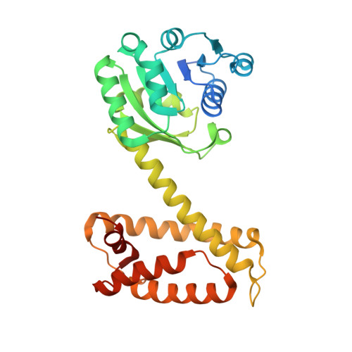
An imine reductase that captures reactive intermediates in the biosynthesis of the indolocarbazole reductasporine.

Daniel-Ivad, P.Ryan, K.S.

(2024) J Biological Chem 300: 105642-105642

  • DOI: https://doi.org/10.1016/j.jbc.2024.105642
  • Primary Citation Related Structures: 
    8U04, 8U05, 8U06, 8U07

  • PubMed Abstract: 

    Imine reductases (IREDs) and reductive aminases have been used in the synthesis of chiral amine products for drug manufacturing; however, little is known about their biological contexts. Here we employ structural studies and site-directed mutagenesis to interrogate the mechanism of the IRED RedE from the biosynthetic pathway to the indolocarbazole natural product reductasporine. Cocrystal structures with the substrate-mimic arcyriaflavin A reveal an extended active site cleft capable of binding two indolocarbazole molecules. Site-directed mutagenesis of a conserved aspartate in the primary binding site reveals a new role for this residue in anchoring the substrate above the NADPH cofactor. Variants targeting the secondary binding site greatly reduce catalytic efficiency, while accumulating oxidized side-products. As indolocarbazole biosynthetic intermediates are susceptible to spontaneous oxidation, we propose the secondary site acts to protect against autooxidation, and the primary site drives catalysis through precise substrate orientation and desolvation effects. The structure of RedE with its extended active site can be the starting point as a new scaffold for engineering IREDs and reductive aminases to intercept large substrates relevant to industrial applications.


  • Organizational Affiliation
    • Department of Chemistry, The University of British Columbia, Vancouver, British Columbia, Canada.

Macromolecule Content 

  • Total Structure Weight: 69.81 kDa 
  • Atom Count: 5,191 
  • Modeled Residue Count: 579 
  • Deposited Residue Count: 630 
  • Unique protein chains: 1

Macromolecules

Find similar proteins by:|  3D Structure
Entity ID: 1
MoleculeChains  Sequence LengthOrganismDetailsImage
RedE
A, B
315uncultured bacteriumMutation(s): 0 
Gene Names: redE
UniProt
Find proteins for A0ACD6B8T4 (uncultured bacterium)
Explore A0ACD6B8T4 
Go to UniProtKB:  A0ACD6B8T4
Entity Groups
Sequence Clusters30% Identity50% Identity70% Identity90% Identity95% Identity100% Identity
UniProt GroupA0ACD6B8T4
Sequence Annotations
Expand
Reference Sequence

Small Molecules

Ligands 6 Unique
IDChains Name / Formula / InChI Key2D Diagram3D Interactions
NAP

Query on NAP


Download:Ideal Coordinates CCD File
C [auth A],
N [auth B]
NADP NICOTINAMIDE-ADENINE-DINUCLEOTIDE PHOSPHATE
C21 H28 N7 O17 P3
XJLXINKUBYWONI-NNYOXOHSSA-N
A7F
(Subject of Investigation/LOI)

Query on A7F


Download:Ideal Coordinates CCD File
D [auth A],
O [auth B]
Arcyriaflavin A
C20 H11 N3 O2
KAJXOWFGKYKMMZ-UHFFFAOYSA-N
IOD

Query on IOD


Download:Ideal Coordinates CCD File
AA [auth B]
BA [auth B]
CA [auth B]
DA [auth B]
EA [auth B]
AA [auth B],
BA [auth B],
CA [auth B],
DA [auth B],
EA [auth B],
FA [auth B],
GA [auth B],
J [auth A],
K [auth A],
L [auth A],
M [auth A]
IODIDE ION
I
XMBWDFGMSWQBCA-UHFFFAOYSA-M
TRS

Query on TRS


Download:Ideal Coordinates CCD File
P [auth B],
Q [auth B]
2-AMINO-2-HYDROXYMETHYL-PROPANE-1,3-DIOL
C4 H12 N O3
LENZDBCJOHFCAS-UHFFFAOYSA-O
EDO

Query on EDO


Download:Ideal Coordinates CCD File
E [auth A],
R [auth B],
S [auth B],
T [auth B],
U [auth B]
1,2-ETHANEDIOL
C2 H6 O2
LYCAIKOWRPUZTN-UHFFFAOYSA-N
CL

Query on CL


Download:Ideal Coordinates CCD File
F [auth A]
G [auth A]
H [auth A]
I [auth A]
V [auth B]
F [auth A],
G [auth A],
H [auth A],
I [auth A],
V [auth B],
W [auth B],
X [auth B],
Y [auth B],
Z [auth B]
CHLORIDE ION
Cl
VEXZGXHMUGYJMC-UHFFFAOYSA-M

Experimental Data & Validation

Experimental Data

  • Method: X-RAY DIFFRACTION
  • Resolution: 1.60 Å
  • R-Value Free:  0.189 (Depositor), 0.190 (DCC) 
  • R-Value Work:  0.155 (Depositor), 0.155 (DCC) 
  • R-Value Observed: 0.156 (Depositor) 
Space Group: P 1 21 1
Unit Cell:
Length ( Å )Angle ( ˚ )
a = 54.114α = 90
b = 110.767β = 107.598
c = 66.987γ = 90
Software Package:
Software NamePurpose
PHENIXrefinement
XDSdata reduction
Aimlessdata scaling
PHASERphasing
Cootmodel building

Structure Validation

View Full Validation Report


Ligand Structure Quality Assessment 


Entry History 

& Funding Information

Deposition Data


Funding OrganizationLocationGrant Number
Natural Sciences and Engineering Research Council (NSERC, Canada)Canada--

Revision History  (Full details and data files)

  • Version 1.0: 2024-09-04
    Type: Initial release
  • Version 1.1: 2025-03-05
    Changes: Database references, Structure summary