8TYG | pdb_00008tyg

Plasmodium vivax PMV-WM08 inhibitor complex


Experimental Data Snapshot

  • Method: X-RAY DIFFRACTION
  • Resolution: 1.64 Å
  • R-Value Free: 
    0.211 (Depositor), 0.211 (DCC) 
  • R-Value Work: 
    0.178 (Depositor), 0.178 (DCC) 
  • R-Value Observed: 
    0.180 (Depositor) 

Starting Model: experimental
View more details

wwPDB Validation   3D Report Full Report


Ligand Structure Quality Assessment 


This is version 1.3 of the entry. See complete history


Literature

Structure-activity analysis of imino-pyrimidinone-fused pyrrolidines aids the development of dual plasmepsin V and plasmepsin X inhibitors.

Hodder, A.N.Sleebs, B.E.Adams, G.Rezazadeh, S.Ngo, A.Jarman, K.Scally, S.Czabotar, P.Wang, H.McCauley, J.A.Olsen, D.B.Cowman, A.F.

(2025) FEBS J 292: 2843-2864

  • DOI: https://doi.org/10.1111/febs.70038
  • Primary Citation of Related Structures:  
    8TYF, 8TYG, 8TYH

  • PubMed Abstract: 

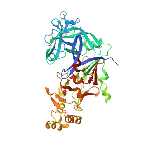
    A library of known aspartic protease inhibitors was screened to identify compounds that inhibit plasmepsin V from Plasmodium vivax. This screen revealed compounds with an imino-pyrimidinone-fused pyrrolidine (IPF) scaffold that exhibited sub-micromolar inhibitory activity against plasmepsin V. Further screening of IPF analogs against the related aspartic protease plasmepsin X showed inhibitory activity, while a third aspartic protease, plasmepsin IX, was not significantly inhibited. Modifications to the P1 biaryl region of the IPF scaffold differentially modulated inhibition of both plasmepsin V and X. Notably, analogs with potent plasmepsin X inhibitory activity successfully blocked the growth of Plasmodium falciparum in vitro. X-ray structures of IPF analogs in complex with plasmepsin V provided insights into their binding mode and revealed avenues to further improve IPF potency and selectivity between plasmepsin V and X. This understanding of how these compounds interact with the active sites of plasmepsin V and X will serve as a foundation for the future design of dual inhibitors targeting these proteases.


  • Organizational Affiliation
    • The Walter and Eliza Hall Institute of Medical Research, Parkville, Australia.

Macromolecules
Find similar proteins by:  (by identity cutoff)  |  3D Structure
Entity ID: 1
MoleculeChains Sequence LengthOrganismDetailsImage
Plasmepsin V461Plasmodium vivax Sal-1Mutation(s): 0 
Gene Names: PMVPVX_116695
EC: 3.4.23
UniProt
Find proteins for A5K302 (Plasmodium vivax (strain Salvador I))
Explore A5K302 
Go to UniProtKB:  A5K302
Entity Groups  
Sequence Clusters30% Identity50% Identity70% Identity90% Identity95% Identity100% Identity
UniProt GroupA5K302
Glycosylation
Glycosylation Sites: 3
Sequence Annotations
Expand
  • Reference Sequence
Oligosaccharides

Help

Entity ID: 2
MoleculeChains Length2D Diagram Glycosylation3D Interactions
2-acetamido-2-deoxy-beta-D-glucopyranose-(1-4)-2-acetamido-2-deoxy-beta-D-glucopyranose
B
2N-Glycosylation
Glycosylation Resources
GlyTouCan:  G42666HT
GlyCosmos:  G42666HT
GlyGen:  G42666HT
Small Molecules
Ligands 4 Unique
IDChains Name / Formula / InChI Key2D Diagram3D Interactions
TEW
Query on TEW

Download Ideal Coordinates CCD File 
D [auth A]6-tungstotellurate(VI)
O24 Te W6
JDLYBIMRCSJRQJ-UHFFFAOYSA-M
SKC (Subject of Investigation/LOI)
Query on SKC

Download Ideal Coordinates CCD File 
C [auth A](2E,4aR,7aS)-6-[3-(4-chlorophenyl)pyridin-2-yl]-7a-(2,5-difluorophenyl)-2-imino-3-methyloctahydro-4H-pyrrolo[3,4-d]pyrimidin-4-one
C24 H20 Cl F2 N5 O
JZQJSLYBFWXABF-YADARESESA-N
NAG
Query on NAG

Download Ideal Coordinates CCD File 
E [auth A],
F [auth A]
2-acetamido-2-deoxy-beta-D-glucopyranose
C8 H15 N O6
OVRNDRQMDRJTHS-FMDGEEDCSA-N
TRS
Query on TRS

Download Ideal Coordinates CCD File 
G [auth A]2-AMINO-2-HYDROXYMETHYL-PROPANE-1,3-DIOL
C4 H12 N O3
LENZDBCJOHFCAS-UHFFFAOYSA-O
Experimental Data & Validation

Experimental Data

  • Method: X-RAY DIFFRACTION
  • Resolution: 1.64 Å
  • R-Value Free:  0.211 (Depositor), 0.211 (DCC) 
  • R-Value Work:  0.178 (Depositor), 0.178 (DCC) 
  • R-Value Observed: 0.180 (Depositor) 
Space Group: I 2 2 2
Unit Cell:
Length ( Å )Angle ( ˚ )
a = 63.772α = 90
b = 86.994β = 90
c = 192.463γ = 90
Software Package:
Software NamePurpose
PHENIXrefinement
XDSdata reduction
Aimlessdata scaling
PHASERphasing

Structure Validation

View Full Validation Report


Ligand Structure Quality Assessment 


Entry History & Funding Information

Deposition Data


Funding OrganizationLocationGrant Number
Wellcome TrustUnited Kingdom--

Revision History  (Full details and data files)

  • Version 1.0: 2024-08-28
    Type: Initial release
  • Version 1.1: 2024-12-25
    Changes: Advisory, Derived calculations, Structure summary
  • Version 1.2: 2025-03-26
    Changes: Database references
  • Version 1.3: 2025-06-18
    Changes: Database references