8TUN | pdb_00008tun

S. thermodepolymerans KpsM-KpsE in Glycolipid 1 state with rigid body fitted KpsT


Experimental Data Snapshot

  • Method: ELECTRON MICROSCOPY
  • Resolution: 3.40 Å
  • Aggregation State: PARTICLE 
  • Reconstruction Method: SINGLE PARTICLE 

wwPDB Validation 3D Report Full Report

Validation slider image for 8TUN

This is version 1.2 of the entry. See complete history

Literature

Molecular insights into capsular polysaccharide secretion.

Kuklewicz, J.Zimmer, J.

(2024) Nature 628: 901-909

  • DOI: https://doi.org/10.1038/s41586-024-07248-9
  • Primary Citation Related Structures: 
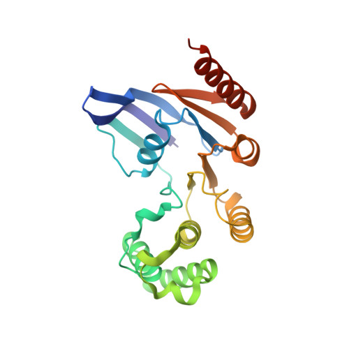
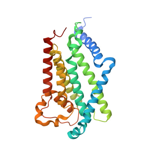
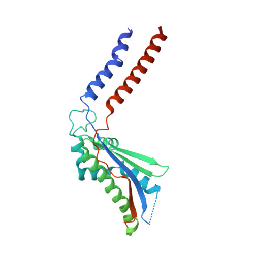
    8TSH, 8TSI, 8TSL, 8TSW, 8TT3, 8TUN

  • PubMed Abstract: 

    Capsular polysaccharides (CPSs) fortify the cell boundaries of many commensal and pathogenic bacteria 1 . Through the ABC-transporter-dependent biosynthesis pathway, CPSs are synthesized intracellularly on a lipid anchor and secreted across the cell envelope by the KpsMT ABC transporter associated with the KpsE and KpsD subunits 1,2 . Here we use structural and functional studies to uncover crucial steps of CPS secretion in Gram-negative bacteria. We show that KpsMT has broad substrate specificity and is sufficient for the translocation of CPSs across the inner bacterial membrane, and we determine the cell surface organization and localization of CPSs using super-resolution fluorescence microscopy. Cryo-electron microscopy analyses of the KpsMT-KpsE complex in six different states reveal a KpsE-encaged ABC transporter, rigid-body conformational rearrangements of KpsMT during ATP hydrolysis and recognition of a glycolipid inside a membrane-exposed electropositive canyon. In vivo CPS secretion assays underscore the functional importance of canyon-lining basic residues. Combined, our analyses suggest a molecular model of CPS secretion by ABC transporters.


  • Organizational Affiliation
    • Department of Molecular Physiology and Biological Physics, University of Virginia School of Medicine, Charlottesville, VA, USA.

Macromolecule Content 

  • Total Structure Weight: 467.33 kDa 
  • Atom Count: 22,046 
  • Modeled Residue Count: 2,725 
  • Deposited Residue Count: 4,136 
  • Unique protein chains: 3

Macromolecules

Find similar proteins by:|  3D Structure
Entity ID: 1
MoleculeChains  Sequence LengthOrganismDetailsImage
ABC transporter ATP-binding protein
A, B
234Caldimonas thermodepolymeransMutation(s): 0 
Gene Names: C1702_11080
Membrane Entity: Yes 
UniProt
Find proteins for A0A2S5T4B3 (Caldimonas thermodepolymerans)
Explore A0A2S5T4B3 
Go to UniProtKB:  A0A2S5T4B3
Entity Groups
Sequence Clusters30% Identity50% Identity70% Identity90% Identity95% Identity100% Identity
UniProt GroupA0A2S5T4B3
Sequence Annotations
Expand
Reference Sequence
Find similar proteins by:|  3D Structure
Entity ID: 2
MoleculeChains  Sequence LengthOrganismDetailsImage
Transport permease protein
C, D
274Caldimonas thermodepolymeransMutation(s): 0 
Gene Names: C1702_11075
Membrane Entity: Yes 
UniProt
Find proteins for A0A2S5T447 (Caldimonas thermodepolymerans)
Explore A0A2S5T447 
Go to UniProtKB:  A0A2S5T447
Entity Groups
Sequence Clusters30% Identity50% Identity70% Identity90% Identity95% Identity100% Identity
UniProt GroupA0A2S5T447
Sequence Annotations
Expand
Reference Sequence
Find similar proteins by:|  3D Structure
Entity ID: 3
MoleculeChains  Sequence LengthOrganismDetailsImage
Capsular biosynthesis protein
E, F, G, H, I
E, F, G, H, I, J, K, L
390Caldimonas thermodepolymeransMutation(s): 0 
Gene Names: C1702_11085
Membrane Entity: Yes 
UniProt
Find proteins for A0A2S5T4A0 (Caldimonas thermodepolymerans)
Explore A0A2S5T4A0 
Go to UniProtKB:  A0A2S5T4A0
Entity Groups
Sequence Clusters30% Identity50% Identity70% Identity90% Identity95% Identity100% Identity
UniProt GroupA0A2S5T4A0
Sequence Annotations
Expand
Reference Sequence

Small Molecules

Ligands 1 Unique
IDChains Name / Formula / InChI Key2D Diagram3D Interactions
KJ9
(Subject of Investigation/LOI)

Query on KJ9


Download:Ideal Coordinates CCD File
M [auth C](2R,5S,8S)-2,5-dihydroxy-5,10-dioxo-8-[(undecanoyloxy)methyl]-4,6,9-trioxa-5lambda~5~-phosphahenicosan-1-yl 3-deoxy-alpha-L-altro-oct-2-ulopyranosidonic acid
C37 H69 O17 P
ATJBCTLIIQRQQQ-KWTBEUQISA-N

Experimental Data & Validation

Experimental Data

  • Method: ELECTRON MICROSCOPY
  • Resolution: 3.40 Å
  • Aggregation State: PARTICLE 
  • Reconstruction Method: SINGLE PARTICLE 

Structure Validation

View Full Validation Report


Entry History 

& Funding Information

Deposition Data


Funding OrganizationLocationGrant Number
National Institutes of Health/National Institute of General Medical Sciences (NIH/NIGMS)United StatesR35GM144130

Revision History  (Full details and data files)

  • Version 1.0: 2024-04-17
    Type: Initial release
  • Version 1.1: 2024-05-08
    Changes: Database references
  • Version 1.2: 2024-10-30
    Changes: Data collection, Structure summary