8TS1 | pdb_00008ts1

Cryo-EM structure of human MRS2 with Mg2+


Experimental Data Snapshot

  • Method: ELECTRON MICROSCOPY
  • Resolution: 2.92 Å
  • Aggregation State: PARTICLE 
  • Reconstruction Method: SINGLE PARTICLE 

wwPDB Validation 3D Report Full Report

Validation slider image for 8TS1

This is version 1.3 of the entry. See complete history

Literature

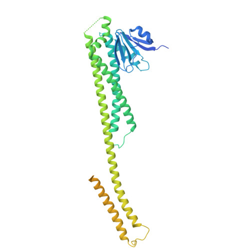
Structure and function of the human mitochondrial MRS2 channel.

He, Z.Tu, Y.C.Tsai, C.W.Mount, J.Zhang, J.Tsai, M.F.Yuan, P.

(2025) Nat Struct Mol Biol 32: 459-468

  • DOI: https://doi.org/10.1038/s41594-024-01420-5
  • Primary Citation Related Structures: 
    8TS1, 8TS2, 8TS3

  • PubMed Abstract: 

    The human mitochondrial RNA splicing 2 protein (MRS2) has been implicated in Mg 2+ transport across mitochondrial inner membranes, thus having an important role in Mg 2+ homeostasis critical for mitochondrial integrity and function. However, the molecular mechanisms underlying its fundamental channel properties such as ion selectivity and regulation remain unclear. Here we present a structural and functional investigation of MRS2. Cryo-electron microscopy structures in various ionic conditions reveal a pentameric channel architecture and the molecular basis of ion permeation and potential regulation mechanisms. Electrophysiological analyses demonstrate that MRS2 is a Ca 2+ -regulated, nonselective channel permeable to Mg 2+ , Ca 2+ , Na + and K + , which contrasts with its prokaryotic ortholog, CorA, operating as a Mg 2+ -gated Mg 2+ channel. Moreover, a conserved arginine ring within the pore of MRS2 functions to restrict cation movements, thus preventing the channel from collapsing the proton motive force that drives mitochondrial adenosine triphosphate synthesis. Together, our results provide a molecular framework for further understanding MRS2 in mitochondrial function and disease.


  • Organizational Affiliation
    • Department of Cell Biology and Physiology, Washington University School of Medicine, Saint Louis, MO, USA.

Macromolecule Content 

  • Total Structure Weight: 277.53 kDa 
  • Atom Count: 12,202 
  • Modeled Residue Count: 1,505 
  • Deposited Residue Count: 2,430 
  • Unique protein chains: 1

Macromolecules

Find similar proteins by:|  3D Structure
Entity ID: 1
MoleculeChains  Sequence LengthOrganismDetailsImage
Magnesium transporter MRS2 homolog, mitochondrial, Soluble cytochrome b562 fusion protein
A, B, C, D, E
486Homo sapiensEscherichia coli
This entity is chimeric
Mutation(s): 0 
Gene Names: MRS2cybC
Membrane Entity: Yes 
UniProt & NIH Common Fund Data Resources
Find proteins for P0ABE7 (Escherichia coli)
Explore P0ABE7 
Go to UniProtKB:  P0ABE7
Find proteins for Q9HD23 (Homo sapiens)
Explore Q9HD23 
Go to UniProtKB:  Q9HD23
PHAROS:  Q9HD23
GTEx:  ENSG00000124532 
Entity Groups
Sequence Clusters30% Identity50% Identity70% Identity90% Identity95% Identity100% Identity
UniProt GroupsP0ABE7Q9HD23
Sequence Annotations
Expand
Reference Sequence

Experimental Data & Validation

Experimental Data

  • Method: ELECTRON MICROSCOPY
  • Resolution: 2.92 Å
  • Aggregation State: PARTICLE 
  • Reconstruction Method: SINGLE PARTICLE 
EM Software:
TaskSoftware PackageVersion
RECONSTRUCTIONcryoSPARC

Structure Validation

View Full Validation Report


Entry History 

& Funding Information

Deposition Data


Funding OrganizationLocationGrant Number
National Institutes of Health/National Institute of Neurological Disorders and Stroke (NIH/NINDS)United StatesR01NS109307

Revision History  (Full details and data files)

  • Version 1.0: 2024-08-14
    Type: Initial release
  • Version 1.1: 2024-10-23
    Changes: Data collection, Database references, Structure summary
  • Version 1.2: 2024-12-11
    Changes: Data collection, Database references
  • Version 1.3: 2025-04-02
    Changes: Data collection, Database references