8TR3 | pdb_00008tr3

Cryo-EM structure of HmAb64 scFv in complex with CNE40 SOSIP trimer


Experimental Data Snapshot

  • Method: ELECTRON MICROSCOPY
  • Resolution: 3.74 Å
  • Aggregation State: PARTICLE 
  • Reconstruction Method: SINGLE PARTICLE 

wwPDB Validation 3D Report Full Report

Validation slider image for 8TR3

This is version 1.1 of the entry. See complete history. 

Literature

Human CD4-binding site antibody elicited by polyvalent DNA prime-protein boost vaccine neutralizes cross-clade tier-2-HIV strains.

Wang, S., Chan, K.W., Wei, D., Ma, X., Liu, S., Hu, G., Park, S., Pan, R., Gu, Y., Nazzari, A.F., Olia, A.S., Xu, K., Lin, B.C., Louder, M.K., McKee, K., Doria-Rose, N.A., Montefiori, D., Seaman, M.S., Zhou, T., Kwong, P.D., Arthos, J., Kong, X.P., Lu, S.

(2024) Nat Commun 15: 4301-4301

  • DOI: https://doi.org/10.1038/s41467-024-48514-8
  • Primary Citation Related Structures: 
    8TR3

  • PubMed Abstract: 

    The vaccine elicitation of HIV tier-2-neutralization antibodies has been a challenge. Here, we report the isolation and characterization of a CD4-binding site (CD4bs) specific monoclonal antibody, HmAb64, from a human volunteer immunized with a polyvalent DNA prime-protein boost HIV vaccine. HmAb64 is derived from heavy chain variable germline gene IGHV1-18 and light chain germline gene IGKV1-39. It has a third heavy chain complementarity-determining region (CDR H3) of 15 amino acids. On a cross-clade panel of 208 HIV-1 pseudo-virus strains, HmAb64 neutralized 20 (10%), including tier-2 strains from clades B, BC, C, and G. The cryo-EM structure of the antigen-binding fragment of HmAb64 in complex with a CNE40 SOSIP trimer revealed details of its recognition; HmAb64 uses both heavy and light CDR3s to recognize the CD4-binding loop, a critical component of the CD4bs. This study demonstrates that a gp120-based vaccine can elicit antibodies capable of tier 2-HIV neutralization.


  • Organizational Affiliation: 
    • Department of Medicine, University of Massachusetts Chan Medical School, Worcester, MA, 01655, USA.

Macromolecule Content 

  • Total Structure Weight: 301.98 kDa 
  • Atom Count: 16,401 
  • Modeled Residue Count: 2,028 
  • Deposited Residue Count: 2,625 
  • Unique protein chains: 4

Macromolecules

Find similar proteins by:|  3D Structure
Entity ID: 1
MoleculeChains  Sequence LengthOrganismDetailsImage
CNE40 SOSIP Envelope glycoprotein gp120A,
E [auth C],
I [auth E]
489Human immunodeficiency virus 1Mutation(s): 4 
Gene Names: env
UniProt
Find proteins for D7S2E5 (Human immunodeficiency virus type 1)
Explore D7S2E5 
Go to UniProtKB:  D7S2E5
Entity Groups
Sequence Clusters30% Identity50% Identity70% Identity90% Identity95% Identity100% Identity
UniProt GroupD7S2E5
Glycosylation
Glycosylation Sites: 11
Sequence Annotations
Expand
Reference Sequence
Find similar proteins by:|  3D Structure
Entity ID: 2
MoleculeChains  Sequence LengthOrganismDetailsImage
CNE40 SOSIP Transmembrane protein gp41B,
F [auth D],
J [auth F]
153Human immunodeficiency virus 1Mutation(s): 2 
Gene Names: env
UniProt
Find proteins for D7S2E5 (Human immunodeficiency virus type 1)
Explore D7S2E5 
Go to UniProtKB:  D7S2E5
Entity Groups
Sequence Clusters30% Identity50% Identity70% Identity90% Identity95% Identity100% Identity
UniProt GroupD7S2E5
Sequence Annotations
Expand
Reference Sequence
Find similar proteins by:|  3D Structure
Entity ID: 3
MoleculeChains  Sequence LengthOrganismDetailsImage
HmAb64 Fv heavy chainC [auth H],
G [auth I],
K [auth J]
124Homo sapiensMutation(s): 0 
Entity Groups
Sequence Clusters30% Identity50% Identity70% Identity90% Identity95% Identity100% Identity
Sequence Annotations
Expand
Reference Sequence
Find similar proteins by:|  3D Structure
Entity ID: 4
MoleculeChains  Sequence LengthOrganismDetailsImage
HmAb64 Fv light chainD [auth L],
H [auth M],
L [auth N]
109Homo sapiensMutation(s): 0 
Entity Groups
Sequence Clusters30% Identity50% Identity70% Identity90% Identity95% Identity100% Identity
Sequence Annotations
Expand
Reference Sequence

Oligosaccharides

Help  
Entity ID: 5
MoleculeChains Length2D Diagram GlycosylationD Interactions
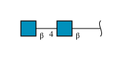
2-acetamido-2-deoxy-beta-D-glucopyranose-(1-4)-2-acetamido-2-deoxy-beta-D-glucopyranose
M [auth P],
N [auth R],
O [auth c],
P [auth e],
Q [auth p],
M [auth P],
N [auth R],
O [auth c],
P [auth e],
Q [auth p],
R [auth r]
2N-Glycosylation
Glycosylation Resources
GlyTouCan: G42666HT
GlyCosmos: G42666HT
GlyGen: G42666HT

Small Molecules

Ligands 1 Unique
IDChains Name / Formula / InChI Key2D Diagram3D Interactions
NAG
(Subject of Investigation/LOI)

Query on NAG


Download:Ideal Coordinates CCD File
AA [auth A]
BA [auth C]
CA [auth C]
DA [auth C]
EA [auth C]
AA [auth A],
BA [auth C],
CA [auth C],
DA [auth C],
EA [auth C],
FA [auth C],
GA [auth C],
HA [auth C],
IA [auth C],
JA [auth C],
KA [auth E],
LA [auth E],
MA [auth E],
NA [auth E],
OA [auth E],
PA [auth E],
QA [auth E],
RA [auth E],
S [auth A],
SA [auth E],
T [auth A],
U [auth A],
V [auth A],
W [auth A],
X [auth A],
Y [auth A],
Z [auth A]
2-acetamido-2-deoxy-beta-D-glucopyranose
C8 H15 N O6
OVRNDRQMDRJTHS-FMDGEEDCSA-N

Experimental Data & Validation

Experimental Data

  • Method: ELECTRON MICROSCOPY
  • Resolution: 3.74 Å
  • Aggregation State: PARTICLE 
  • Reconstruction Method: SINGLE PARTICLE 
EM Software:
TaskSoftware PackageVersion
RECONSTRUCTIONcryoSPARC3.3.2
MODEL REFINEMENTPHENIX1.20.1-4487

Structure Validation

View Full Validation Report


Entry History 

& Funding Information

Deposition Data


Funding OrganizationLocationGrant Number
National Institutes of Health/National Institute Of Allergy and Infectious Diseases (NIH/NIAID)United States--

Revision History  (Full details and data files)

  • Version 1.0: 2024-06-19
    Type: Initial release
  • Version 1.1: 2024-11-06
    Changes: Data collection, Structure summary