8TP8

Structure of the C. crescentus WYL-activator, DriD, bound to ssDNA and cognate DNA


Experimental Data Snapshot

  • Method: X-RAY DIFFRACTION
  • Resolution: 2.74 Å
  • R-Value Free: 0.241 
  • R-Value Work: 0.196 
  • R-Value Observed: 0.198 

wwPDB Validation   3D Report Full Report


This is version 1.3 of the entry. See complete history


Literature

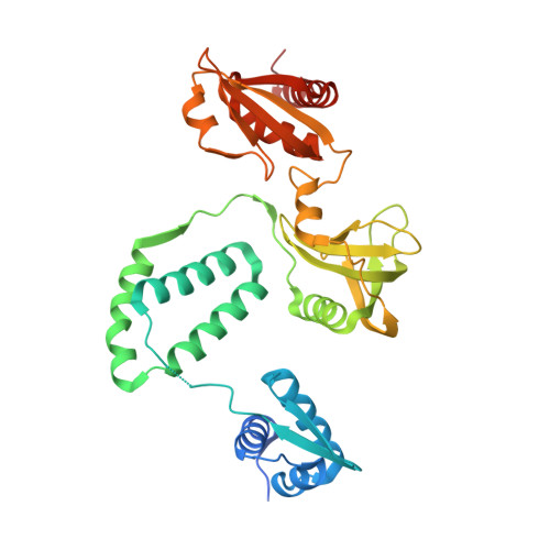
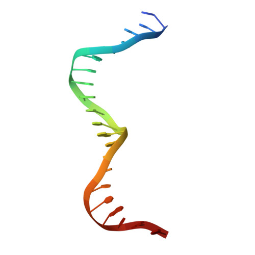
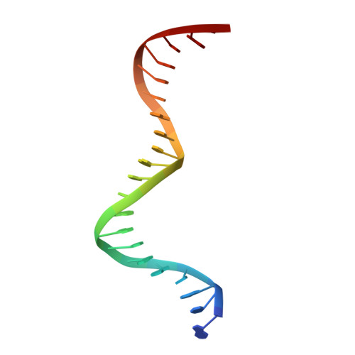
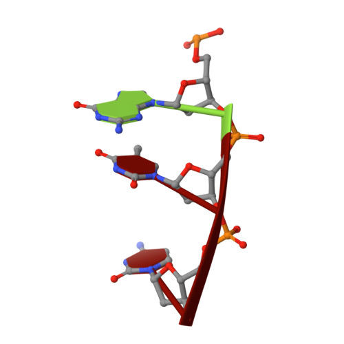
Structure of the WYL-domain containing transcription activator, DriD, in complex with ssDNA effector and DNA target site.

Schumacher, M.A.Cannistraci, E.Salinas, R.Lloyd, D.Messner, E.Gozzi, K.

(2024) Nucleic Acids Res 52: 1435-1449

  • DOI: https://doi.org/10.1093/nar/gkad1198
  • Primary Citation of Related Structures:  
    8TP8, 8TPK

  • PubMed Abstract: 

    Transcription regulators play central roles in orchestrating responses to changing environmental conditions. Recently the Caulobacter crescentus transcription activator DriD, which belongs to the newly defined WYL-domain family, was shown to regulate DNA damage responses independent of the canonical SOS pathway. However, the molecular mechanisms by which DriD and other WYL-regulators sense environmental signals and recognize DNA are not well understood. We showed DriD DNA-binding is triggered by its interaction with ssDNA, which is produced during DNA damage. Here we describe the structure of the full-length C. crescentus DriD bound to both target DNA and effector ssDNA. DriD consists of an N-terminal winged-HTH (wHTH) domain, linker region, three-helix bundle, WYL-domain and C-terminal WCX-dimer domain. Strikingly, DriD binds DNA using a novel, asymmetric DNA-binding mechanism that results from different conformations adopted by the linker. Although the linker does not touch DNA, our data show that contacts it makes with the wHTH are key for specific DNA binding. The structure indicates how ssDNA-effector binding to the WYL-domain impacts wHTH DNA binding. In conclusion, we present the first structure of a WYL-activator bound to both effector and target DNA. The structure unveils a unique, asymmetric DNA binding mode that is likely conserved among WYL-activators.


  • Organizational Affiliation

    Department of Biochemistry, 307 Research Dr., Box 3711, Duke University Medical Center, Durham, NC 27710, USA.


Macromolecules

Find similar proteins by:  (by identity cutoff)  |  3D Structure
Entity ID: 1
MoleculeChains Sequence LengthOrganismDetailsImage
DeoR-family transcriptional regulatorA,
B,
G [auth C],
H [auth D]
347Caulobacter vibrioides NA1000Mutation(s): 0 
Gene Names: CCNA_01151
UniProt
Find proteins for A0A0H3C5Q6 (Caulobacter vibrioides (strain NA1000 / CB15N))
Explore A0A0H3C5Q6 
Go to UniProtKB:  A0A0H3C5Q6
Entity Groups  
Sequence Clusters30% Identity50% Identity70% Identity90% Identity95% Identity100% Identity
UniProt GroupA0A0H3C5Q6
Sequence Annotations
Expand
  • Reference Sequence

Find similar nucleic acids by:  Sequence   |   3D Structure  

Entity ID: 2
MoleculeChains LengthOrganismImage
DNA (5'-D(*AP*TP*AP*CP*GP*AP*CP*AP*GP*TP*TP*AP*CP*TP*GP*TP*CP*GP*TP*AP*T)-3')C [auth U],
K [auth F]
21Caulobacter vibrioides
Sequence Annotations
Expand
  • Reference Sequence

Find similar nucleic acids by:  Sequence   |   3D Structure  

Entity ID: 3
MoleculeChains LengthOrganismImage
DNA (5'-D(*AP*TP*AP*CP*GP*AP*CP*AP*GP*TP*AP*AP*CP*TP*GP*TP*CP*GP*TP*AP*T)-3')D [auth R],
L [auth T]
21Caulobacter vibrioides
Sequence Annotations
Expand
  • Reference Sequence

Find similar nucleic acids by:  Sequence   |   3D Structure  

Entity ID: 4
MoleculeChains LengthOrganismImage
DNA (5'-D(P*GP*TP*C)-3')E [auth L],
F [auth Y],
I [auth J],
J [auth K]
3Caulobacter vibrioides
Sequence Annotations
Expand
  • Reference Sequence
Experimental Data & Validation

Experimental Data

  • Method: X-RAY DIFFRACTION
  • Resolution: 2.74 Å
  • R-Value Free: 0.241 
  • R-Value Work: 0.196 
  • R-Value Observed: 0.198 
  • Space Group: P 1 21 1
Unit Cell:
Length ( Å )Angle ( ˚ )
a = 69.26α = 90
b = 178.941β = 95.24
c = 92.15γ = 90
Software Package:
Software NamePurpose
PHENIXrefinement
SCALAdata scaling
XDSdata reduction
MOLREPphasing

Structure Validation

View Full Validation Report


Entry History & Funding Information

Deposition Data


Funding OrganizationLocationGrant Number
National Institutes of Health/National Institute of General Medical Sciences (NIH/NIGMS)United StatesR35GM130290

Revision History  (Full details and data files)

  • Version 1.0: 2023-11-29
    Type: Initial release
  • Version 1.1: 2024-01-10
    Changes: Database references
  • Version 1.2: 2024-02-21
    Changes: Database references
  • Version 1.3: 2024-03-13
    Changes: Source and taxonomy