8TOX | pdb_00008tox

Cryo-EM structure of BG505 Env mutant A517E in complex with antibody ACS202 Fab


Experimental Data Snapshot

  • Method: ELECTRON MICROSCOPY
  • Resolution: 2.30 Å
  • Aggregation State: PARTICLE 
  • Reconstruction Method: SINGLE PARTICLE 

wwPDB Validation 3D Report Full Report

Validation slider image for 8TOX

This is version 1.2 of the entry. See complete history. 

Literature

Vaccine-elicited and naturally elicited antibodies differ in their recognition of the HIV-1 fusion peptide.

Reveiz, M., Xu, K., Lee, M., Wang, S., Olia, A.S., Harris, D.R., Liu, K., Liu, T., Schaub, A.J., Stephens, T., Wang, Y., Zhang, B., Huang, R., Tsybovsky, Y., Kwong, P.D., Rawi, R.

(2024) Front Immunol 15: 1484029-1484029

  • DOI: https://doi.org/10.3389/fimmu.2024.1484029
  • Primary Citation Related Structures: 
    8TOX

  • PubMed Abstract: 

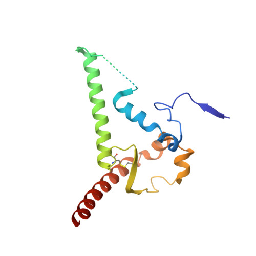
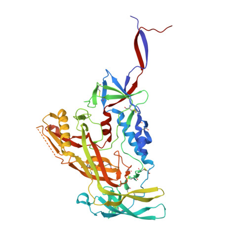
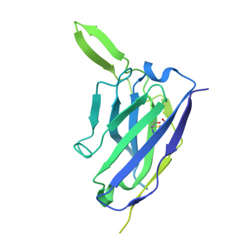
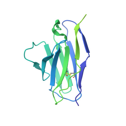
    Broadly neutralizing antibodies have been proposed as templates for HIV-1 vaccine design, but it has been unclear how similar vaccine-elicited antibodies are to their naturally elicited templates. To provide insight, here we compare the recognition of naturally elicited and vaccine-elicited antibodies targeting the HIV-1 fusion peptide, which comprises envelope (Env) residues 512-526, with the most common sequence being AVGIGAVFLGFLGAA. Naturally elicited antibodies bound peptides with substitutions to negatively charged amino acids at residue positions 517-520 substantially better than the most common sequence, despite these substitutions rarely appearing in HIV-1; by contrast, vaccine-elicited antibodies were less tolerant of sequence variation, with no substitution of residues 512-516 showing increased binding. Molecular dynamics analysis and cryo-EM structural analysis of the naturally elicited ACS202 antibody in complex with the HIV-1 Env trimer with an alanine 517 to glutamine substitution suggested enhanced binding to result from electrostatic interactions with positively charged antibody residues. Overall, vaccine-elicited antibodies appeared to be more fully optimized to bind the most common fusion peptide sequence, perhaps reflecting the immunization with fusion peptide of the vaccine-elicited antibodies.


  • Organizational Affiliation: 
    • Vaccine Research Center, National Institute of Allergy and Infectious Diseases, National Institutes of Health, Bethesda, MD, United States.

Macromolecule Content 

  • Total Structure Weight: 389.08 kDa 
  • Atom Count: 22,008 
  • Modeled Residue Count: 2,490 
  • Deposited Residue Count: 3,234 
  • Unique protein chains: 4

Macromolecules

Find similar proteins by:|  3D Structure
Entity ID: 1
MoleculeChains  Sequence LengthOrganismDetailsImage
Envelope glycoprotein gp41A [auth B],
C [auth A],
E [auth F]
153Human immunodeficiency virus 1Mutation(s): 12 
Gene Names: env
UniProt
Find proteins for Q2N0S6 (Human immunodeficiency virus type 1)
Explore Q2N0S6 
Go to UniProtKB:  Q2N0S6
Entity Groups
Sequence Clusters30% Identity50% Identity70% Identity90% Identity95% Identity100% Identity
UniProt GroupQ2N0S6
Glycosylation
Glycosylation Sites: 3
Sequence Annotations
Expand
Reference Sequence
Find similar proteins by:|  3D Structure
Entity ID: 2
MoleculeChains  Sequence LengthOrganismDetailsImage
Envelope glycoprotein gp120B [auth G],
D [auth C],
F [auth I]
475Human immunodeficiency virus 1Mutation(s): 12 
Gene Names: env
UniProt
Find proteins for Q2N0S6 (Human immunodeficiency virus type 1)
Explore Q2N0S6 
Go to UniProtKB:  Q2N0S6
Entity Groups
Sequence Clusters30% Identity50% Identity70% Identity90% Identity95% Identity100% Identity
UniProt GroupQ2N0S6
Glycosylation
Glycosylation Sites: 18
Sequence Annotations
Expand
Reference Sequence
Find similar proteins by:|  3D Structure
Entity ID: 3
MoleculeChains  Sequence LengthOrganismDetailsImage
antibody ACS202 Fab heavy chainG [auth H],
I [auth D],
K [auth J]
236Homo sapiensMutation(s): 0 
Entity Groups
Sequence Clusters30% Identity50% Identity70% Identity90% Identity95% Identity100% Identity
Sequence Annotations
Expand
Reference Sequence
Find similar proteins by:|  3D Structure
Entity ID: 4
MoleculeChains  Sequence LengthOrganismDetailsImage
antibody ACS202 Fab light chainH [auth L],
J [auth E],
L [auth K]
214Homo sapiensMutation(s): 0 
Entity Groups
Sequence Clusters30% Identity50% Identity70% Identity90% Identity95% Identity100% Identity
Sequence Annotations
Expand
Reference Sequence

Oligosaccharides

Help  
Entity ID: 5
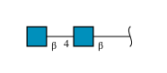
MoleculeChains Length2D Diagram GlycosylationD Interactions
2-acetamido-2-deoxy-beta-D-glucopyranose-(1-4)-2-acetamido-2-deoxy-beta-D-glucopyranose
AB [auth 0],
BA [auth b],
BB [auth 1],
DA [auth d],
EA [auth e],
AB [auth 0],
BA [auth b],
BB [auth 1],
DA [auth d],
EA [auth e],
EB [auth 4],
FB [auth 5],
GA [auth g],
GB [auth 6],
HB [auth 7],
IA [auth i],
JA [auth j],
JB [auth 9],
KA [auth k],
M,
N,
NA [auth n],
OA [auth o],
P,
PA [auth p],
QA [auth q],
R,
S,
SA [auth s],
T,
UA [auth u],
VA [auth v],
W,
X,
XA [auth x],
Y,
Z,
ZA [auth z]
2N-Glycosylation
Glycosylation Resources
GlyTouCan: G42666HT
GlyCosmos: G42666HT
GlyGen: G42666HT
Entity ID: 6
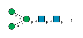
MoleculeChains Length2D Diagram GlycosylationD Interactions
alpha-D-mannopyranose-(1-3)-[alpha-D-mannopyranose-(1-6)]beta-D-mannopyranose-(1-4)-2-acetamido-2-deoxy-beta-D-glucopyranose-(1-4)-2-acetamido-2-deoxy-beta-D-glucopyranose
CA [auth c],
DB [auth 3],
FA [auth f],
KB [auth AA],
MA [auth m],
CA [auth c],
DB [auth 3],
FA [auth f],
KB [auth AA],
MA [auth m],
O,
TA [auth t],
V,
WA [auth w]
5N-Glycosylation
Glycosylation Resources
GlyTouCan: G22768VO
GlyCosmos: G22768VO
GlyGen: G22768VO
Entity ID: 7
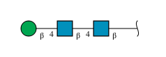
MoleculeChains Length2D Diagram GlycosylationD Interactions
beta-D-mannopyranose-(1-4)-2-acetamido-2-deoxy-beta-D-glucopyranose-(1-4)-2-acetamido-2-deoxy-beta-D-glucopyranose
AA [auth a],
HA [auth h],
IB [auth 8],
Q,
RA [auth r],
AA [auth a],
HA [auth h],
IB [auth 8],
Q,
RA [auth r],
YA [auth y]
3N-Glycosylation
Glycosylation Resources
GlyTouCan: G15407YE
GlyCosmos: G15407YE
GlyGen: G15407YE
Entity ID: 8
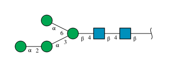
MoleculeChains Length2D Diagram GlycosylationD Interactions
alpha-D-mannopyranose-(1-2)-alpha-D-mannopyranose-(1-3)-[alpha-D-mannopyranose-(1-6)]beta-D-mannopyranose-(1-4)-2-acetamido-2-deoxy-beta-D-glucopyranose-(1-4)-2-acetamido-2-deoxy-beta-D-glucopyranoseCB [auth 2],
LA [auth l],
U
6N-Glycosylation
Glycosylation Resources
GlyTouCan: G56014GC
GlyCosmos: G56014GC
GlyGen: G56014GC

Small Molecules

Ligands 1 Unique
IDChains Name / Formula / InChI Key2D Diagram3D Interactions
NAG
(Subject of Investigation/LOI)

Query on NAG


Download:Ideal Coordinates CCD File
LB [auth B]
MB [auth G]
NB [auth G]
OB [auth G]
PB [auth A]
LB [auth B],
MB [auth G],
NB [auth G],
OB [auth G],
PB [auth A],
QB [auth C],
RB [auth C],
SB [auth C],
TB [auth F],
UB [auth I],
VB [auth I],
WB [auth I]
2-acetamido-2-deoxy-beta-D-glucopyranose
C8 H15 N O6
OVRNDRQMDRJTHS-FMDGEEDCSA-N

Experimental Data & Validation

Experimental Data

  • Method: ELECTRON MICROSCOPY
  • Resolution: 2.30 Å
  • Aggregation State: PARTICLE 
  • Reconstruction Method: SINGLE PARTICLE 
EM Software:
TaskSoftware PackageVersion
RECONSTRUCTIONcryoSPARC3.3
MODEL REFINEMENTPHENIX1.20

Structure Validation

View Full Validation Report


Entry History 

& Funding Information

Deposition Data


Funding OrganizationLocationGrant Number
National Institutes of Health/National Institute Of Allergy and Infectious Diseases (NIH/NIAID)United States--

Revision History  (Full details and data files)

  • Version 1.0: 2024-08-14
    Type: Initial release
  • Version 1.1: 2024-10-23
    Changes: Data collection, Structure summary
  • Version 1.2: 2025-02-26
    Changes: Data collection, Database references