8TI7

Crystal structure of profilin from Dermatophagoides pteronyssinus in complex with a poly(L-proline) peptide


Experimental Data Snapshot

  • Method: X-RAY DIFFRACTION
  • Resolution: 2.40 Å
  • R-Value Free: 0.195 
  • R-Value Work: 0.166 

wwPDB Validation   3D Report Full Report


This is version 1.0 of the entry. See complete history


Literature

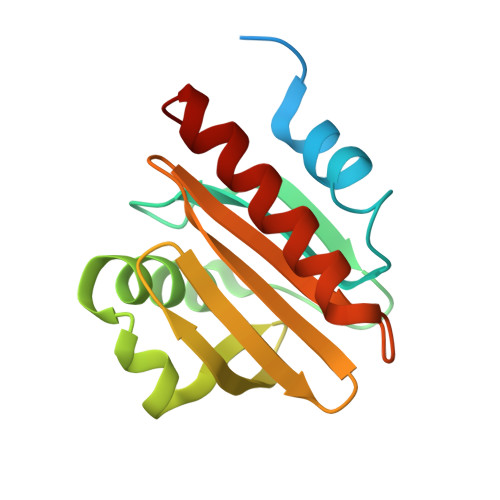
Structural homology of mite profilins to plant profilins is not indicative of allergic cross-reactivity.

O'Malley, A.Sankaran, S.Carriuolo, A.Khatri, K.Kowal, K.Chruszcz, M.

(2024) Biol Chem 

  • DOI: https://doi.org/10.1515/hsz-2023-0366
  • Primary Citation of Related Structures:  
    8TI5, 8TI6, 8TI7, 8V5Y

  • PubMed Abstract: 

    Structural and allergenic characterization of mite profilins has not been previously pursued to a similar extent as plant profilins. Here, we describe structures of profilins originating from Tyrophagus putrescentiae (registered allergen Tyr p 36.0101) and Dermatophagoides pteronyssinus (here termed Der p profilin), which are the first structures of profilins from Arachnida. Additionally, the thermal stabilities of mite and plant profilins are compared, suggesting that the high number of cysteine residues in mite profilins may play a role in their increased stability. We also examine the cross-reactivity of plant and mite profilins as well as investigate the relevance of these profilins in mite inhalant allergy. Despite their high structural similarity to other profilins, mite profilins have low sequence identity with plant and human profilins. Subsequently, these mite profilins most likely do not display cross-reactivity with plant profilins. At the same time the profilins have highly conserved poly(l-proline) and actin binding sites.


  • Organizational Affiliation

    Department of Biochemistry and Molecular Biology, 3078 Michigan State University , 603 Wilson Road, East Lansing, MI 48824, USA.


Macromolecules
Find similar proteins by:  (by identity cutoff)  |  3D Structure
Entity ID: 1
MoleculeChains Sequence LengthOrganismDetailsImage
Profilin154Dermatophagoides pteronyssinusMutation(s): 0 
Gene Names: LOC113791125
UniProt
Find proteins for A0A2L0EBJ5 (Dermatophagoides pteronyssinus)
Explore A0A2L0EBJ5 
Go to UniProtKB:  A0A2L0EBJ5
Entity Groups  
Sequence Clusters30% Identity50% Identity70% Identity90% Identity95% Identity100% Identity
UniProt GroupA0A2L0EBJ5
Sequence Annotations
Expand
  • Reference Sequence

Find similar proteins by:  Sequence   |   3D Structure  

Entity ID: 2
MoleculeChains Sequence LengthOrganismDetailsImage
poly(L-proline)12synthetic constructMutation(s): 0 
Entity Groups  
Sequence Clusters30% Identity50% Identity70% Identity90% Identity95% Identity100% Identity
Sequence Annotations
Expand
  • Reference Sequence
Small Molecules
Ligands 1 Unique
IDChains Name / Formula / InChI Key2D Diagram3D Interactions
SO4
Query on SO4

Download Ideal Coordinates CCD File 
AA [auth O]
BA [auth O]
Q [auth A]
R [auth A]
S [auth A]
AA [auth O],
BA [auth O],
Q [auth A],
R [auth A],
S [auth A],
T [auth G],
U [auth I],
V [auth I],
W [auth K],
X [auth K],
Y [auth M],
Z [auth M]
SULFATE ION
O4 S
QAOWNCQODCNURD-UHFFFAOYSA-L
Experimental Data & Validation

Experimental Data

  • Method: X-RAY DIFFRACTION
  • Resolution: 2.40 Å
  • R-Value Free: 0.195 
  • R-Value Work: 0.166 
  • Space Group: H 3
Unit Cell:
Length ( Å )Angle ( ˚ )
a = 152.802α = 90
b = 152.802β = 90
c = 186.854γ = 120
Software Package:
Software NamePurpose
REFMACrefinement
HKL-3000data reduction
HKL-3000data scaling
MOLREPphasing

Structure Validation

View Full Validation Report


Entry History & Funding Information

Deposition Data


Funding OrganizationLocationGrant Number
National Institutes of Health/National Institute Of Allergy and Infectious Diseases (NIH/NIAID)United StatesR01AI077653

Revision History  (Full details and data files)

  • Version 1.0: 2024-05-08
    Type: Initial release