8TD0

Structure of PYCR1 complexed with 5-oxo-7a-phenyl-hexahydropyrrolo[2,1-b][1,3]thiazole-3-carboxylic acid


Experimental Data Snapshot

  • Method: X-RAY DIFFRACTION
  • Resolution: 2.20 Å
  • R-Value Free: 0.259 
  • R-Value Work: 0.222 
  • R-Value Observed: 0.224 

wwPDB Validation   3D Report Full Report


Ligand Structure Quality Assessment 


This is version 1.1 of the entry. See complete history


Literature

Novel Fragment Inhibitors of PYCR1 from Docking-Guided X-ray Crystallography.

Meeks, K.R.Ji, J.Protopopov, M.V.Tarkhanova, O.O.Moroz, Y.S.Tanner, J.J.

(2024) J Chem Inf Model 64: 1704-1718

  • DOI: https://doi.org/10.1021/acs.jcim.3c01879
  • Primary Citation of Related Structures:  
    8TCU, 8TCV, 8TCW, 8TCX, 8TCY, 8TCZ, 8TD0, 8TD1

  • PubMed Abstract: 

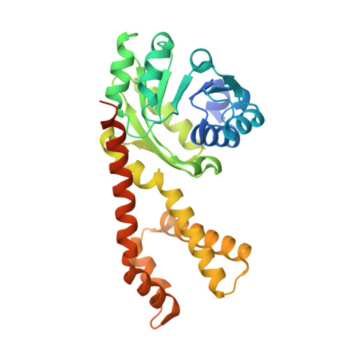
    The proline biosynthetic enzyme Δ 1 -pyrroline-5-carboxylate (P5C) reductase 1 (PYCR1) is one of the most consistently upregulated enzymes across multiple cancer types and central to the metabolic rewiring of cancer cells. Herein, we describe a fragment-based, structure-first approach to the discovery of PYCR1 inhibitors. Thirty-seven fragment-like carboxylic acids in the molecular weight range of 143-289 Da were selected from docking and then screened using X-ray crystallography as the primary assay. Strong electron density was observed for eight compounds, corresponding to a crystallographic hit rate of 22%. The fragments are novel compared to existing proline analog inhibitors in that they block both the P5C substrate pocket and the NAD(P)H binding site. Four hits showed inhibition of PYCR1 in kinetic assays, and one has lower apparent IC 50 than the current best proline analog inhibitor. These results show proof-of-concept for our inhibitor discovery approach and provide a basis for fragment-to-lead optimization.


  • Organizational Affiliation

    Department of Biochemistry, University of Missouri, Columbia, Missouri 65211, United States.


Macromolecules
Find similar proteins by:  (by identity cutoff)  |  3D Structure
Entity ID: 1
MoleculeChains Sequence LengthOrganismDetailsImage
Pyrroline-5-carboxylate reductase 1, mitochondrial
A, B, C, D, E
316Homo sapiensMutation(s): 0 
Gene Names: PYCR1
EC: 1.5.1.2
UniProt & NIH Common Fund Data Resources
Find proteins for P32322 (Homo sapiens)
Explore P32322 
Go to UniProtKB:  P32322
PHAROS:  P32322
GTEx:  ENSG00000183010 
Entity Groups  
Sequence Clusters30% Identity50% Identity70% Identity90% Identity95% Identity100% Identity
UniProt GroupP32322
Sequence Annotations
Expand
  • Reference Sequence
Experimental Data & Validation

Experimental Data

  • Method: X-RAY DIFFRACTION
  • Resolution: 2.20 Å
  • R-Value Free: 0.259 
  • R-Value Work: 0.222 
  • R-Value Observed: 0.224 
  • Space Group: C 1 2 1
Unit Cell:
Length ( Å )Angle ( ˚ )
a = 109.349α = 90
b = 179.119β = 106.83
c = 87.914γ = 90
Software Package:
Software NamePurpose
PHENIXrefinement
Aimlessdata scaling
XDSdata reduction
PHENIXphasing

Structure Validation

View Full Validation Report


Ligand Structure Quality Assessment 


Entry History & Funding Information

Deposition Data


Funding OrganizationLocationGrant Number
National Institutes of Health/National Institute of General Medical Sciences (NIH/NIGMS)United States1R01GM132640

Revision History  (Full details and data files)

  • Version 1.0: 2024-03-06
    Type: Initial release
  • Version 1.1: 2024-03-20
    Changes: Database references