8TB8

Sequence specific (AATT) orientation of Hoechst molecules at a unique minor groove binding site (position1) within a self-assembled 3D DNA lattice (4x5)


Experimental Data Snapshot

  • Method: X-RAY DIFFRACTION
  • Resolution: 2.95 Å
  • R-Value Free: 0.272 
  • R-Value Work: 0.235 
  • R-Value Observed: 0.237 

wwPDB Validation   3D Report Full Report


Ligand Structure Quality Assessment 


This is version 1.0 of the entry. See complete history


Literature

Site-Specific Arrangement and Structure Determination of Minor Groove Binding Molecules in Self-Assembled Three-Dimensional DNA Crystals.

Simmons, C.R.Buchberger, A.Henry, S.J.W.Novacek, A.Fahmi, N.E.MacCulloch, T.Stephanopoulos, N.Yan, H.

(2023) J Am Chem Soc 145: 26075-26085

  • DOI: https://doi.org/10.1021/jacs.3c07802
  • Primary Citation of Related Structures:  
    8T7B, 8T7X, 8T80, 8TA8, 8TA9, 8TAJ, 8TAM, 8TAP, 8TAQ, 8TB3, 8TB4, 8TB8, 8TBD, 8TBO, 8TC2, 8TC4, 8TC6, 8TDT

  • PubMed Abstract: 

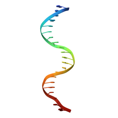
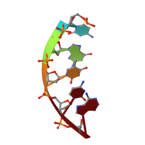
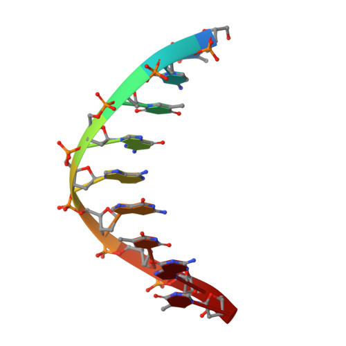
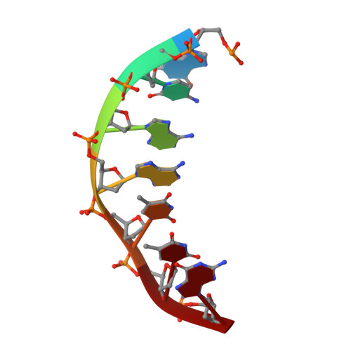
    The structural analysis of guest molecules in rationally designed and self-assembling DNA crystals has proven an elusive goal since its conception. Oligonucleotide frameworks provide an especially attractive route toward studying DNA-binding molecules by using three-dimensional lattices with defined sequence and structure. In this work, we site-specifically position a suite of minor groove binding molecules, and solve their structures via X-ray crystallography as a proof-of-principle toward scaffolding larger guest species. Two crystal motifs were used to precisely immobilize the molecules DAPI, Hoechst, and netropsin at defined positions in the lattice, allowing us to control occupancy within the crystal. We also solved the structure of a three-ring imidazole-pyrrole-pyrrole polyamide molecule, which sequence-specifically packs in an antiparallel dimeric arrangement within the minor groove. Finally, we engineered a crystal designed to position both netropsin and the polyamide at two distinct locations within the same lattice. Our work elucidates the design principles for the spatial arrangement of functional guests within lattices and opens new potential opportunities for the use of DNA crystals to display and structurally characterize small molecules, peptides, and ultimately proteins of unknown structure.


  • Organizational Affiliation

    Biodesign Center for Molecular Design and Biomimetics, Arizona State University 1001 S. McAllister Ave., Tempe, Arizona 85287, United States.


Macromolecules

Find similar nucleic acids by:  Sequence   |   3D Structure  

Entity ID: 1
MoleculeChains LengthOrganismImage
DNA (5'-D(*GP*AP*CP*AP*AP*TP*TP*GP*CP*TP*GP*AP*CP*GP*AP*CP*AP*CP*TP*CP*A*(HT1))-3')21synthetic construct
Sequence Annotations
Expand
  • Reference Sequence

Find similar nucleic acids by:  Sequence   |   3D Structure  

Entity ID: 2
MoleculeChains LengthOrganismImage
DNA (5'-D(P*CP*GP*TP*CP*A)-3')5synthetic construct
Sequence Annotations
Expand
  • Reference Sequence

Find similar nucleic acids by:  Sequence   |   3D Structure  

Entity ID: 3
MoleculeChains LengthOrganismImage
DNA (5'-D(*TP*CP*TP*GP*AP*GP*TP*GP*T)-3')9synthetic construct
Sequence Annotations
Expand
  • Reference Sequence

Find similar nucleic acids by:  Sequence   |   3D Structure  

Entity ID: 4
MoleculeChains LengthOrganismImage
DNA (5'-D(P*GP*CP*AP*AP*TP*TP*G)-3')7synthetic construct
Sequence Annotations
Expand
  • Reference Sequence
Small Molecules
Ligands 2 Unique
IDChains Name / Formula / InChI Key2D Diagram3D Interactions
HT1
Query on HT1

Download Ideal Coordinates CCD File 
F [auth D]2'-(4-ETHOXYPHENYL)-5-(4-METHYL-1-PIPERAZINYL)-2,5'-BI-BENZIMIDAZOLE
C27 H28 N6 O
PRDFBSVERLRRMY-UHFFFAOYSA-N
MG
Query on MG

Download Ideal Coordinates CCD File 
E [auth C]MAGNESIUM ION
Mg
JLVVSXFLKOJNIY-UHFFFAOYSA-N
Experimental Data & Validation

Experimental Data

  • Method: X-RAY DIFFRACTION
  • Resolution: 2.95 Å
  • R-Value Free: 0.272 
  • R-Value Work: 0.235 
  • R-Value Observed: 0.237 
  • Space Group: P 32 2 1
Unit Cell:
Length ( Å )Angle ( ˚ )
a = 68.195α = 90
b = 68.195β = 90
c = 60.661γ = 120
Software Package:
Software NamePurpose
PHENIXrefinement
HKL-2000data scaling
HKL-2000data reduction
PHASERphasing

Structure Validation

View Full Validation Report


Ligand Structure Quality Assessment 


Entry History & Funding Information

Deposition Data


Funding OrganizationLocationGrant Number
National Science Foundation (NSF, United States)United States1360635
National Institutes of Health/National Institute of General Medical Sciences (NIH/NIGMS)United StatesR01GM104960
National Science Foundation (NSF, United States)United StatesNSF2004250

Revision History  (Full details and data files)

  • Version 1.0: 2023-12-20
    Type: Initial release