8T8I

Structure of VHH-Fab complex with engineered Elbow FNQIKG, Crystal Kappa and SER substitutions


Experimental Data Snapshot

  • Method: X-RAY DIFFRACTION
  • Resolution: 2.52 Å
  • R-Value Free: 0.243 
  • R-Value Work: 0.191 
  • R-Value Observed: 0.194 

wwPDB Validation   3D Report Full Report


Ligand Structure Quality Assessment 


This is version 1.1 of the entry. See complete history


Literature

Engineered antigen-binding fragments for enhanced crystallization of antibody:antigen complexes.

Bruce, H.A.Singer, A.U.Filippova, E.V.Blazer, L.L.Adams, J.J.Enderle, L.Ben-David, M.Radley, E.H.Mao, D.Y.L.Pau, V.Orlicky, S.Sicheri, F.Kurinov, I.Atwell, S.Kossiakoff, A.A.Sidhu, S.S.

(2024) Protein Sci 33: e4824-e4824

  • DOI: https://doi.org/10.1002/pro.4824
  • Primary Citation of Related Structures:  
    8T58, 8T6I, 8T7F, 8T7G, 8T7I, 8T8I, 8T9Y, 8TRS, 8TRT, 8TS5

  • PubMed Abstract: 

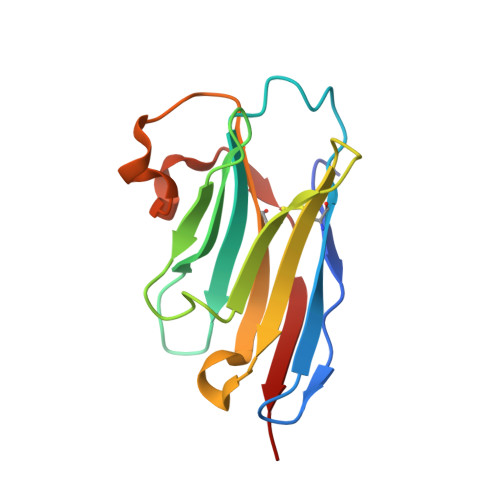
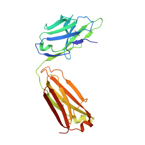
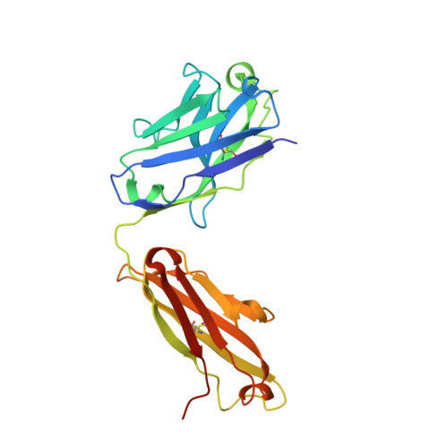
    The atomic-resolution structural information that X-ray crystallography can provide on the binding interface between a Fab and its cognate antigen is highly valuable for understanding the mechanism of interaction. However, many Fab:antigen complexes are recalcitrant to crystallization, making the endeavor a considerable effort with no guarantee of success. Consequently, there have been significant steps taken to increase the likelihood of Fab:antigen complex crystallization by altering the Fab framework. In this investigation, we applied the surface entropy reduction strategy coupled with phage-display technology to identify a set of surface substitutions that improve the propensity of a human Fab framework to crystallize. In addition, we showed that combining these surface substitutions with previously reported Crystal Kappa and elbow substitutions results in an extraordinary improvement in Fab and Fab:antigen complex crystallizability, revealing a strong synergistic relationship between these sets of substitutions. Through comprehensive Fab and Fab:antigen complex crystallization screenings followed by structure determination and analysis, we defined the roles that each of these substitutions play in facilitating crystallization and how they complement each other in the process.


  • Organizational Affiliation

    School of Pharmacy, University of Waterloo, Waterloo, Canada.


Macromolecules
Find similar proteins by:  (by identity cutoff)  |  3D Structure
Entity ID: 1
MoleculeChains Sequence LengthOrganismDetailsImage
VHH domain129Homo sapiensMutation(s): 0 
Entity Groups  
Sequence Clusters30% Identity50% Identity70% Identity90% Identity95% Identity100% Identity
Sequence Annotations
Expand
  • Reference Sequence
Find similar proteins by:  (by identity cutoff)  |  3D Structure
Entity ID: 2
MoleculeChains Sequence LengthOrganismDetailsImage
Fab light chainB [auth L]213Homo sapiensMutation(s): 0 
Entity Groups  
Sequence Clusters30% Identity50% Identity70% Identity90% Identity95% Identity100% Identity
Sequence Annotations
Expand
  • Reference Sequence
Find similar proteins by:  (by identity cutoff)  |  3D Structure
Entity ID: 3
MoleculeChains Sequence LengthOrganismDetailsImage
Fab heavy chainC [auth H]238Homo sapiensMutation(s): 0 
Entity Groups  
Sequence Clusters30% Identity50% Identity70% Identity90% Identity95% Identity100% Identity
Sequence Annotations
Expand
  • Reference Sequence
Small Molecules
Ligands 4 Unique
IDChains Name / Formula / InChI Key2D Diagram3D Interactions
SIN (Subject of Investigation/LOI)
Query on SIN

Download Ideal Coordinates CCD File 
D [auth A]SUCCINIC ACID
C4 H6 O4
KDYFGRWQOYBRFD-UHFFFAOYSA-N
PEG (Subject of Investigation/LOI)
Query on PEG

Download Ideal Coordinates CCD File 
E [auth A]DI(HYDROXYETHYL)ETHER
C4 H10 O3
MTHSVFCYNBDYFN-UHFFFAOYSA-N
PO4 (Subject of Investigation/LOI)
Query on PO4

Download Ideal Coordinates CCD File 
F [auth L],
G [auth L]
PHOSPHATE ION
O4 P
NBIIXXVUZAFLBC-UHFFFAOYSA-K
CL (Subject of Investigation/LOI)
Query on CL

Download Ideal Coordinates CCD File 
H,
I [auth H]
CHLORIDE ION
Cl
VEXZGXHMUGYJMC-UHFFFAOYSA-M
Experimental Data & Validation

Experimental Data

  • Method: X-RAY DIFFRACTION
  • Resolution: 2.52 Å
  • R-Value Free: 0.243 
  • R-Value Work: 0.191 
  • R-Value Observed: 0.194 
  • Space Group: P 21 21 21
Unit Cell:
Length ( Å )Angle ( ˚ )
a = 52.221α = 90
b = 72.771β = 90
c = 242.398γ = 90
Software Package:
Software NamePurpose
REFMACrefinement
XDSdata reduction
Aimlessdata scaling
PHASERphasing
PDB_EXTRACTdata extraction

Structure Validation

View Full Validation Report


Ligand Structure Quality Assessment 


Entry History & Funding Information

Deposition Data


Funding OrganizationLocationGrant Number
National Institutes of Health/National Institute of General Medical Sciences (NIH/NIGMS)United States--

Revision History  (Full details and data files)

  • Version 1.0: 2023-11-29
    Type: Initial release
  • Version 1.1: 2024-01-03
    Changes: Database references