8T8A | pdb_00008t8a

Structure of arginine oxidase from Pseudomonas sp. TRU 7192


Experimental Data Snapshot

  • Method: X-RAY DIFFRACTION
  • Resolution: 3.40 Å
  • R-Value Free: 
    0.313 (Depositor), 0.320 (DCC) 
  • R-Value Work: 
    0.212 (Depositor), 0.240 (DCC) 
  • R-Value Observed: 
    0.217 (Depositor) 

Starting Model: in silico
View more details

wwPDB Validation   3D Report Full Report


Ligand Structure Quality Assessment 


This is version 1.3 of the entry. See complete history


Literature

Open and closed structures of L-arginine oxidase by cryo-electron microscopy and X-ray crystallography.

Yamaguchi, H.Takahashi, K.Numoto, N.Suzuki, H.Tatsumi, M.Kamegawa, A.Nishikawa, K.Asano, Y.Mizukoshi, T.Miyano, H.Fujiyoshi, Y.Sugiki, M.

(2025) J Biochem 177: 27-36

  • DOI: https://doi.org/10.1093/jb/mvae070
  • Primary Citation of Related Structures:  
    8JT7, 8T8A

  • PubMed Abstract: 

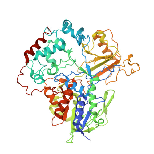
    L-arginine oxidase (AROD, EC 1.4.3.25) is an oxidoreductase that catalyzes the deamination of L-arginine, with flavin adenine dinucleotide (FAD) as a cofactor. Recently identified AROD from Pseudomonas sp. TPU 7192 (PT-AROD) demonstrates high selectivity for L-arginine. This enzyme is useful for accurate assays of L-arginine in biological samples. The structural characteristics of the FAD-dependent AROD, however, remain unknown. Here, we report the structure of PT-AROD at a resolution of 2.3 Å by cryo-electron microscopy. PT-AROD adopts an octameric structure with D4 symmetry, which is consistent with its molecular weight in solution, estimated by mass photometry. Comparative analysis of this structure with that determined using X-ray crystallography reveals open and closed forms of the lid-like loop at the entrance to the substrate pocket. Furthermore, mutation of Glu493, located at the substrate binding site, diminishes substrate selectivity, suggesting that this residue contributes significantly to the high selectivity of PT-AROD.


  • Organizational Affiliation
    • Research Institute for Bioscience Products & Fine Chemicals, Ajinomoto Co., Inc. 1-1 Suzuki-cho, Kawasaki-ku, Kawasaki 210-8681, Japan.

Macromolecules
Find similar proteins by:  (by identity cutoff)  |  3D Structure
Entity ID: 1
MoleculeChains Sequence LengthOrganismDetailsImage
Amine oxidoreductase
A, B, C, D
596Pseudomonas sp.Mutation(s): 0 
Gene Names: AYO28_23740
EC: 1.13.12.3
UniProt
Find proteins for A0A177SH44 (Pseudomonas putida)
Explore A0A177SH44 
Go to UniProtKB:  A0A177SH44
Entity Groups  
Sequence Clusters30% Identity50% Identity70% Identity90% Identity95% Identity100% Identity
UniProt GroupA0A177SH44
Sequence Annotations
Expand
  • Reference Sequence
Experimental Data & Validation

Experimental Data

  • Method: X-RAY DIFFRACTION
  • Resolution: 3.40 Å
  • R-Value Free:  0.313 (Depositor), 0.320 (DCC) 
  • R-Value Work:  0.212 (Depositor), 0.240 (DCC) 
  • R-Value Observed: 0.217 (Depositor) 
Space Group: C 2 2 21
Unit Cell:
Length ( Å )Angle ( ˚ )
a = 168.274α = 90
b = 200.219β = 90
c = 168.542γ = 90
Software Package:
Software NamePurpose
REFMACrefinement
XDSdata reduction
Aimlessdata scaling
PHASERphasing

Structure Validation

View Full Validation Report


Ligand Structure Quality Assessment 


Entry History & Funding Information

Deposition Data


Funding OrganizationLocationGrant Number
Not funded--

Revision History  (Full details and data files)

  • Version 1.0: 2024-06-26
    Type: Initial release
  • Version 1.1: 2024-10-23
    Changes: Structure summary
  • Version 1.2: 2025-01-08
    Changes: Database references
  • Version 1.3: 2025-01-15
    Changes: Database references