8T56 | pdb_00008t56

Structure of mechanically activated ion channel OSCA1.2 in peptidiscs


Experimental Data Snapshot

  • Method: ELECTRON MICROSCOPY
  • Resolution: 2.80 Å
  • Aggregation State: PARTICLE 
  • Reconstruction Method: SINGLE PARTICLE 

Starting Model: experimental
View more details

wwPDB Validation 3D Report Full Report

Validation slider image for 8T56

This is version 1.1 of the entry. See complete history

Literature

Structure of mechanically activated ion channel OSCA2.3 reveals mobile elements in the transmembrane domain.

Jojoa-Cruz, S.Burendei, B.Lee, W.H.Ward, A.B.

(2024) Structure 32: 157

  • DOI: https://doi.org/10.1016/j.str.2023.11.009
  • Primary Citation Related Structures: 
    8T56, 8T57

  • PubMed Abstract: 

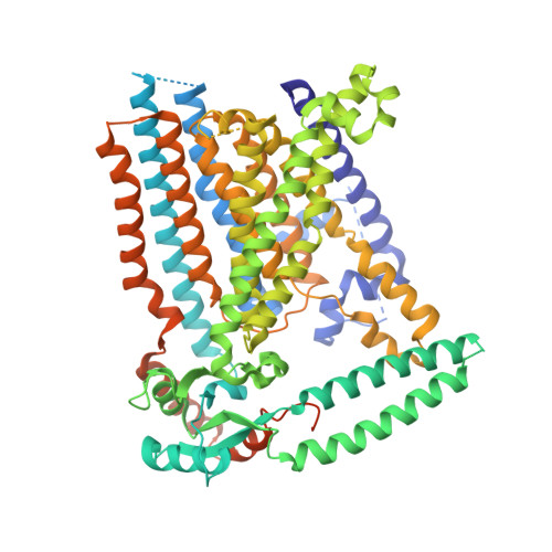
    Members of the OSCA/TMEM63 family are mechanically activated ion channels and structures of some OSCA members have revealed the architecture of these channels and structural features that are potentially involved in mechanosensation. However, these structures are all in a similar state and information about the motion of different elements of the structure is limited, preventing a deeper understanding of how these channels work. Here, we used cryoelectron microscopy to determine high-resolution structures of Arabidopsis thaliana OSCA1.2 and OSCA2.3 in peptidiscs. The structure of OSCA1.2 matches previous structures of the same protein in different environments. Yet, in OSCA2.3, the TM6a-TM7 linker adopts a different conformation that constricts the pore on its cytoplasmic side. Furthermore, coevolutionary sequence analysis uncovered a conserved interaction between the TM6a-TM7 linker and the beam-like domain (BLD). Our results reveal conformational heterogeneity and differences in conserved interactions between the TMD and BLD among members of the OSCA family.


  • Organizational Affiliation
    • Department of Integrative Structural and Computational Biology, The Scripps Research Institute, La Jolla, CA 92037, USA.

Macromolecule Content 

  • Total Structure Weight: 225.4 kDa 
  • Atom Count: 13,096 
  • Modeled Residue Count: 1,498 
  • Deposited Residue Count: 1,858 
  • Unique protein chains: 2

Macromolecules

Find similar proteins by:|  3D Structure
Entity ID: 1
MoleculeChains  Sequence LengthOrganismDetailsImage
Calcium permeable stress-gated cation channel 1
A, B
781Arabidopsis thalianaMutation(s): 0 
Gene Names: CSC1At4g22120F1N20.220
Membrane Entity: Yes 
UniProt
Find proteins for Q5XEZ5 (Arabidopsis thaliana)
Explore Q5XEZ5 
Go to UniProtKB:  Q5XEZ5
Entity Groups
Sequence Clusters30% Identity50% Identity70% Identity90% Identity95% Identity100% Identity
UniProt GroupQ5XEZ5
Sequence Annotations
Expand
Reference Sequence
Find similar proteins by:|  3D Structure
Entity ID: 2
MoleculeChains  Sequence LengthOrganismDetailsImage
NSPr peptide
C, D, E, F, G
C, D, E, F, G, H, I, J
37synthetic constructMutation(s): 0 

Small Molecules

Ligands 4 Unique
IDChains Name / Formula / InChI Key2D Diagram3D Interactions
LBN

Query on LBN


Download:Ideal Coordinates CCD File
HA [auth B],
V [auth A]
1-palmitoyl-2-oleoyl-sn-glycero-3-phosphocholine
C42 H82 N O8 P
WTJKGGKOPKCXLL-VYOBOKEXSA-N
Y01

Query on Y01


Download:Ideal Coordinates CCD File
K [auth A],
W [auth B]
CHOLESTEROL HEMISUCCINATE
C31 H50 O4
WLNARFZDISHUGS-MIXBDBMTSA-N
CLR

Query on CLR


Download:Ideal Coordinates CCD File
IA [auth C]
JA [auth D]
MA [auth E]
OA [auth G]
PA [auth H]
IA [auth C],
JA [auth D],
MA [auth E],
OA [auth G],
PA [auth H],
SA [auth I]
CHOLESTEROL
C27 H46 O
HVYWMOMLDIMFJA-DPAQBDIFSA-N
PLM

Query on PLM


Download:Ideal Coordinates CCD File
AA [auth B]
BA [auth B]
CA [auth B]
DA [auth B]
EA [auth B]
AA [auth B],
BA [auth B],
CA [auth B],
DA [auth B],
EA [auth B],
FA [auth B],
GA [auth B],
KA [auth D],
L [auth A],
LA [auth D],
M [auth A],
N [auth A],
NA [auth E],
O [auth A],
P [auth A],
Q [auth A],
QA [auth H],
R [auth A],
RA [auth H],
S [auth A],
T [auth A],
TA [auth I],
U [auth A],
X [auth B],
Y [auth B],
Z [auth B]
PALMITIC ACID
C16 H32 O2
IPCSVZSSVZVIGE-UHFFFAOYSA-N

Experimental Data & Validation

Experimental Data

  • Method: ELECTRON MICROSCOPY
  • Resolution: 2.80 Å
  • Aggregation State: PARTICLE 
  • Reconstruction Method: SINGLE PARTICLE 
EM Software:
TaskSoftware PackageVersion
RECONSTRUCTIONRELION3.1
MODEL REFINEMENTCoot0.9
MODEL REFINEMENTPHENIX1.18.2-3874
MODEL REFINEMENTRosetta3.10

Structure Validation

View Full Validation Report


Entry History 

& Funding Information

Deposition Data


Funding OrganizationLocationGrant Number
National Institutes of Health/National Heart, Lung, and Blood Institute (NIH/NHLBI)United StatesHL143297

Revision History  (Full details and data files)

  • Version 1.0: 2023-12-27
    Type: Initial release
  • Version 1.1: 2024-02-14
    Changes: Database references