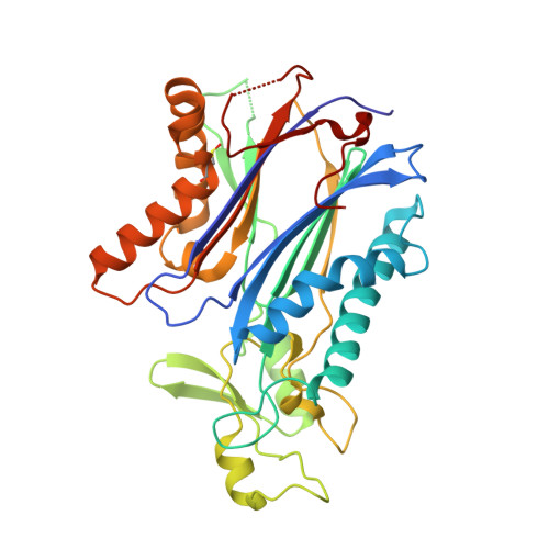
8T2J

Structure of the catalytic domain of PPM1D/Wip1 serine/threonine phosphatase


Experimental Data Snapshot

  • Method: X-RAY DIFFRACTION
  • Resolution: 1.80 Å
  • R-Value Free: 0.219 
  • R-Value Work: 0.175 
  • R-Value Observed: 0.176 

Starting Model: experimental
View more details

wwPDB Validation   3D Report Full Report


This is version 1.3 of the entry. See complete history


Literature

Crystal structure and mechanistic studies of the PPM1D serine/threonine phosphatase catalytic domain.

Kumar, J.P.Kosek, D.Durell, S.R.Miller Jenkins, L.M.Debnath, S.Coussens, N.P.Hall, M.D.Appella, D.H.Dyda, F.Mazur, S.J.Appella, E.

(2024) J Biol Chem 300: 107561-107561

  • DOI: https://doi.org/10.1016/j.jbc.2024.107561
  • Primary Citation of Related Structures:  
    8T2J

  • PubMed Abstract: 

    Protein phosphatase 1D (PPM1D, Wip1) is induced by the tumor suppressor p53 during DNA damage response signaling and acts as an oncoprotein in several human cancers. Although PPM1D is a potential therapeutic target, insights into its atomic structure were challenging due to flexible regions unique to this family member. Here we report the first crystal structure of the PPM1D catalytic domain to 1.8 Å resolution. The structure reveals the active site with two Mg 2+ ions bound, similar to other structures. The flap subdomain and B-loop, which are crucial for substrate recognition and catalysis, were also resolved, with the flap forming two short helices and three short β-strands that are followed by an irregular loop. Unexpectedly, a nitrogen-oxygen-sulfur bridge was identified in the catalytic domain. Molecular dynamics simulations and kinetic studies provided further mechanistic insights into the regulation of PPM1D catalytic activity. In particular, the kinetic experiments demonstrated a magnesium concentration-dependent lag in PPM1D attaining steady-state velocity, a feature of hysteretic enzymes that show slow transitions compared with catalytic turnover. All combined, these results advance the understanding of PPM1D function and will support the development of PPM1D-targeted therapeutics.


  • Organizational Affiliation

    Laboratory of Cell Biology, NCI, National Institutes of Health, Bethesda, MD 20892, United States.


Macromolecules
Find similar proteins by:  (by identity cutoff)  |  3D Structure
Entity ID: 1
MoleculeChains Sequence LengthOrganismDetailsImage
Protein phosphatase 1D345Homo sapiensMutation(s): 5 
Gene Names: PPM1DWIP1
EC: 3.1.3.16
UniProt & NIH Common Fund Data Resources
Find proteins for O15297 (Homo sapiens)
Explore O15297 
Go to UniProtKB:  O15297
PHAROS:  O15297
GTEx:  ENSG00000170836 
Entity Groups  
Sequence Clusters30% Identity50% Identity70% Identity90% Identity95% Identity100% Identity
UniProt GroupO15297
Sequence Annotations
Expand
  • Reference Sequence
Experimental Data & Validation

Experimental Data

  • Method: X-RAY DIFFRACTION
  • Resolution: 1.80 Å
  • R-Value Free: 0.219 
  • R-Value Work: 0.175 
  • R-Value Observed: 0.176 
  • Space Group: P 1 21 1
Unit Cell:
Length ( Å )Angle ( ˚ )
a = 49.664α = 90
b = 59.93β = 92.13
c = 49.566γ = 90
Software Package:
Software NamePurpose
PHENIXrefinement
XSCALEdata scaling
XDSdata reduction
PHASERphasing

Structure Validation

View Full Validation Report


Entry History & Funding Information

Deposition Data


Funding OrganizationLocationGrant Number
National Institutes of Health/National Institute of Diabetes and Digestive and Kidney Disease (NIH/NIDDK)United StatesResearch resulting in this structure was funded in part by the Intramural Program of the NIDDK

Revision History  (Full details and data files)

  • Version 1.0: 2024-06-12
    Type: Initial release
  • Version 1.1: 2024-07-31
    Changes: Database references
  • Version 1.2: 2024-08-14
    Changes: Database references
  • Version 1.3: 2024-10-23
    Changes: Structure summary