8T1G | pdb_00008t1g

The crystal structure of hemagglutinin form a h7n9 influenza virus (a/shanghai/1/2013) in complex with antibody 1E11


Experimental Data Snapshot

  • Method: X-RAY DIFFRACTION
  • Resolution: 3.50 Å
  • R-Value Free: 
    0.215 (Depositor), 0.215 (DCC) 
  • R-Value Work: 
    0.182 (Depositor), 0.182 (DCC) 
  • R-Value Observed: 
    0.182 (Depositor) 

Starting Model: experimental
View more details

wwPDB Validation 3D Report Full Report

Validation slider image for 8T1G

Ligand Structure Quality Assessment 


This is version 1.4 of the entry. See complete history

Literature

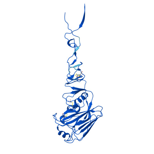
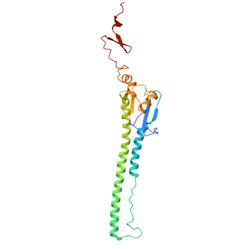
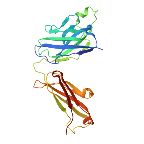
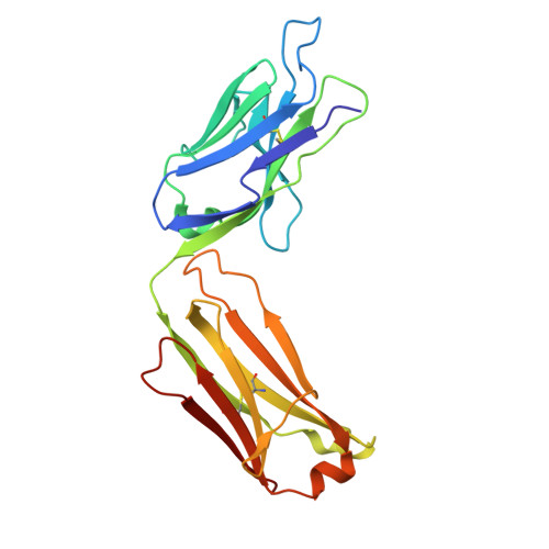
Distinct binding modes drive the broad neutralization profile of two persistent influenza hemagglutinin stem-specific antibody lineages.

Mantus, G.E.Cerutti, G.Chambers, M.Gillespie, R.A.Shimberg, G.D.Spangler, A.Gorman, J.Zhou, T.Shen, C.H.Kanekiyo, M.Kwong, P.D.Shapiro, L.Andrews, S.F.

(2025) Structure 33: 869

  • DOI: https://doi.org/10.1016/j.str.2025.02.010
  • Primary Citation Related Structures: 
    8T1G, 8VEB, 8VED, 8VEE, 8VEF

  • PubMed Abstract: 

    Elicitation of antibodies to the influenza hemagglutinin stem is a critical part of universal influenza vaccine strategies. While numerous broadly reactive stem antibodies have been isolated, our understanding of how these antibodies mature within the human B cell repertoire is limited. Here, we isolated and tracked two stem-specific antibody lineages over a decade in a single participant that received multiple seasonal and pandemic influenza vaccinations. Despite similar binding and neutralization profiles, antibodies from these lineages utilized fundamentally different interactions to engage the central epitope on the influenza stem. Structural analysis of an unmutated common ancestor from one lineage identified critical residues that were the main drivers of increased affinity and breadth to group 1 influenza subtypes. These observations demonstrate the heterogeneous pathways by which stem-specific antibodies can mature within the human B cell repertoire.


  • Organizational Affiliation
    • Vaccine Research Center, National Institute of Allergy and Infectious Diseases, National Institutes of Health, Bethesda, MD, USA.

Macromolecule Content 

  • Total Structure Weight: 328.82 kDa 
  • Atom Count: 22,809 
  • Modeled Residue Count: 2,914 
  • Deposited Residue Count: 2,946 
  • Unique protein chains: 4

Macromolecules

Find similar proteins by:|  3D Structure
Entity ID: 1
MoleculeChains  Sequence LengthOrganismDetailsImage
Hemagglutinin HA1
A, C, E
321Influenza A virusMutation(s): 0 
Gene Names: HA
UniProt
Find proteins for R4NN21 (Influenza A virus)
Explore R4NN21 
Go to UniProtKB:  R4NN21
Entity Groups
Sequence Clusters30% Identity50% Identity70% Identity90% Identity95% Identity100% Identity
UniProt GroupR4NN21
Glycosylation
Glycosylation Sites: 2
Sequence Annotations
Expand
Reference Sequence
Find similar proteins by:|  3D Structure
Entity ID: 2
MoleculeChains  Sequence LengthOrganismDetailsImage
Hemagglutinin HA2
B, D, F
210Influenza A virusMutation(s): 0 
Gene Names: HA
UniProt
Find proteins for R4NN21 (Influenza A virus)
Explore R4NN21 
Go to UniProtKB:  R4NN21
Entity Groups
Sequence Clusters30% Identity50% Identity70% Identity90% Identity95% Identity100% Identity
UniProt GroupR4NN21
Glycosylation
Glycosylation Sites: 2
Sequence Annotations
Expand
Reference Sequence
Find similar proteins by:|  3D Structure
Entity ID: 3
MoleculeChains  Sequence LengthOrganismDetailsImage
1E11 Fab Heavy chain
G, H, J
232Homo sapiensMutation(s): 0 
Find similar proteins by:|  3D Structure
Entity ID: 4
MoleculeChains  Sequence LengthOrganismDetailsImage
1E11 Fab Light chain
I, K, L
219Homo sapiensMutation(s): 0 

Oligosaccharides

Help  
Entity ID: 5
MoleculeChains Length2D Diagram GlycosylationD Interactions
beta-D-mannopyranose-(1-4)-2-acetamido-2-deoxy-beta-D-glucopyranose-(1-4)-2-acetamido-2-deoxy-beta-D-glucopyranose
M, O, Q
3N-Glycosylation
Glycosylation Resources
GlyTouCan: G15407YE
GlyCosmos: G15407YE
GlyGen: G15407YE
Entity ID: 6
MoleculeChains Length2D Diagram GlycosylationD Interactions
alpha-D-mannopyranose-(1-3)-beta-D-mannopyranose-(1-4)-2-acetamido-2-deoxy-beta-D-glucopyranose-(1-4)-2-acetamido-2-deoxy-beta-D-glucopyranose
N, R
4N-Glycosylation
Glycosylation Resources
GlyTouCan: G81315DD
GlyCosmos: G81315DD
GlyGen: G81315DD
Entity ID: 7
MoleculeChains Length2D Diagram GlycosylationD Interactions
alpha-D-mannopyranose-(1-6)-beta-D-mannopyranose-(1-4)-2-acetamido-2-deoxy-beta-D-glucopyranose-(1-4)-2-acetamido-2-deoxy-beta-D-glucopyranose
P
4N-Glycosylation
Glycosylation Resources
GlyTouCan: G22573RC
GlyCosmos: G22573RC
GlyGen: G22573RC

Small Molecules

Ligands 2 Unique
IDChains Name / Formula / InChI Key2D Diagram3D Interactions
NAG

Query on NAG


Download:Ideal Coordinates CCD File
AA [auth F]
T [auth A]
U [auth B]
W [auth C]
X [auth D]
AA [auth F],
T [auth A],
U [auth B],
W [auth C],
X [auth D],
Z [auth E]
2-acetamido-2-deoxy-beta-D-glucopyranose
C8 H15 N O6
OVRNDRQMDRJTHS-FMDGEEDCSA-N
CA

Query on CA


Download:Ideal Coordinates CCD File
S [auth A],
V [auth C],
Y [auth E]
CALCIUM ION
Ca
BHPQYMZQTOCNFJ-UHFFFAOYSA-N

Experimental Data & Validation

Experimental Data

  • Method: X-RAY DIFFRACTION
  • Resolution: 3.50 Å
  • R-Value Free:  0.215 (Depositor), 0.215 (DCC) 
  • R-Value Work:  0.182 (Depositor), 0.182 (DCC) 
  • R-Value Observed: 0.182 (Depositor) 
Space Group: P 42 21 2
Unit Cell:
Length ( Å )Angle ( ˚ )
a = 303.277α = 90
b = 303.277β = 90
c = 153.461γ = 90
Software Package:
Software NamePurpose
PHENIXrefinement
SCALEPACKdata scaling
HKL-2000data reduction
PHASERphasing

Structure Validation

View Full Validation Report


Ligand Structure Quality Assessment 


Entry History 

& Funding Information

Deposition Data


Funding OrganizationLocationGrant Number
National Institutes of Health/National Institute Of Allergy and Infectious Diseases (NIH/NIAID)United States--

Revision History  (Full details and data files)

  • Version 1.0: 2024-05-29
    Type: Initial release
  • Version 1.1: 2024-10-09
    Changes: Structure summary
  • Version 1.2: 2025-03-26
    Changes: Database references
  • Version 1.3: 2025-04-02
    Changes: Database references
  • Version 1.4: 2025-05-14
    Changes: Database references