


Crystal structure of Yersinia pestis dihydrofolate reductase in complex with Trimethoprim
Shaw, G.X., Cherry, S., Tropea, J.E., Waugh, D.S., Ji, X.To be published.
Experimental Data Snapshot
Starting Model: experimental
View more details
Entity ID: 1 | |||||
|---|---|---|---|---|---|
| Molecule | Chains | Sequence Length | Organism | Details | Image |
| Dihydrofolate reductase | 164 | Yersinia pestis | Mutation(s): 0 Gene Names: folA, tmrA, YPO0486 EC: 1.5.1.3 | 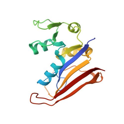 | |
UniProt | |||||
Entity Groups | |||||
| Sequence Clusters | 30% Identity50% Identity70% Identity90% Identity95% Identity100% Identity | ||||
| UniProt Group | A0A3N4BLI0 | ||||
Sequence AnnotationsExpand | |||||
Reference Sequence | |||||
| Ligands 4 Unique | |||||
|---|---|---|---|---|---|
| ID | Chains | Name / Formula / InChI Key | 2D Diagram | 3D Interactions | |
| TOP (Subject of Investigation/LOI) Download:Ideal Coordinates CCD File | C [auth A], K [auth B] | TRIMETHOPRIM C14 H18 N4 O3 IEDVJHCEMCRBQM-UHFFFAOYSA-N |  | ||
| SO4 Download:Ideal Coordinates CCD File | D [auth A] E [auth A] F [auth A] L [auth B] M [auth B] | SULFATE ION O4 S QAOWNCQODCNURD-UHFFFAOYSA-L |  | ||
| EDO Download:Ideal Coordinates CCD File | G [auth A] | 1,2-ETHANEDIOL C2 H6 O2 LYCAIKOWRPUZTN-UHFFFAOYSA-N |  | ||
| CL Download:Ideal Coordinates CCD File | H [auth A], I [auth A], J [auth A], P [auth B] | CHLORIDE ION Cl VEXZGXHMUGYJMC-UHFFFAOYSA-M |  | ||
| Length ( Å ) | Angle ( ˚ ) |
|---|---|
| a = 115.421 | α = 90 |
| b = 115.421 | β = 90 |
| c = 146.325 | γ = 120 |
| Software Name | Purpose |
|---|---|
| PHENIX | refinement |
| SCALEPACK | data scaling |
| DENZO | data reduction |
| PHASER | phasing |
| Funding Organization | Location | Grant Number |
|---|---|---|
| National Institutes of Health/National Cancer Institute (NIH/NCI) | United States | Intramural Research Program |


