8SUR

TMEM16F bound with Niclosamide


Experimental Data Snapshot

  • Method: ELECTRON MICROSCOPY
  • Resolution: 3.10 Å
  • Aggregation State: CELL 
  • Reconstruction Method: SINGLE PARTICLE 

wwPDB Validation   3D Report Full Report


This is version 2.0 of the entry. See complete history


Literature

Identification of a drug binding pocket in TMEM16F calcium-activated ion channel and lipid scramblase.

Feng, S.Puchades, C.Ko, J.Wu, H.Chen, Y.Figueroa, E.E.Gu, S.Han, T.W.Ho, B.Cheng, T.Li, J.Shoichet, B.Jan, Y.N.Cheng, Y.Jan, L.Y.

(2023) Nat Commun 14: 4874-4874

  • DOI: https://doi.org/10.1038/s41467-023-40410-x
  • Primary Citation of Related Structures:  
    8SUN, 8SUR, 8TAG, 8TAI, 8TAL

  • PubMed Abstract: 

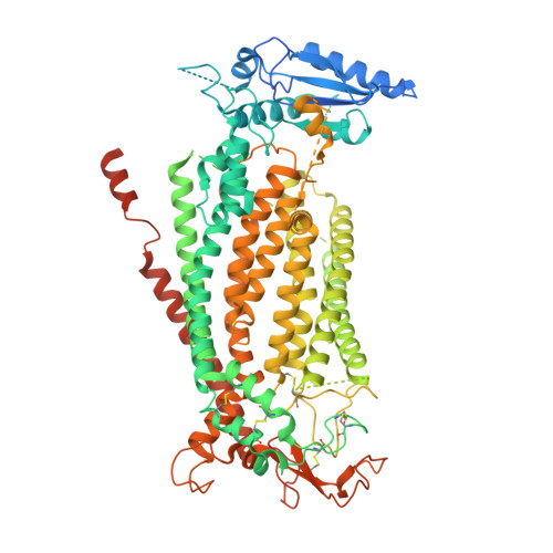
    The dual functions of TMEM16F as Ca 2+ -activated ion channel and lipid scramblase raise intriguing questions regarding their molecular basis. Intrigued by the ability of the FDA-approved drug niclosamide to inhibit TMEM16F-dependent syncytia formation induced by SARS-CoV-2, we examined cryo-EM structures of TMEM16F with or without bound niclosamide or 1PBC, a known blocker of TMEM16A Ca 2+ -activated Cl - channel. Here, we report evidence for a lipid scrambling pathway along a groove harboring a lipid trail outside the ion permeation pore. This groove contains the binding pocket for niclosamide and 1PBC. Mutations of two residues in this groove specifically affect lipid scrambling. Whereas mutations of some residues in the binding pocket of niclosamide and 1PBC reduce their inhibition of TMEM16F-mediated Ca 2+ influx and PS exposure, other mutations preferentially affect the ability of niclosamide and/or 1PBC to inhibit TMEM16F-mediated PS exposure, providing further support for separate pathways for ion permeation and lipid scrambling.


  • Organizational Affiliation

    Department of Physiology, University of California San Francisco (UCSF) School of Medicine, San Francisco, CA, USA.


Macromolecules
Find similar proteins by:  (by identity cutoff)  |  3D Structure
Entity ID: 1
MoleculeChains Sequence LengthOrganismDetailsImage
Anoctamin-6
A, B
911Mus musculusMutation(s): 0 
Gene Names: Ano6Tmem16f
Membrane Entity: Yes 
UniProt & NIH Common Fund Data Resources
Find proteins for Q6P9J9 (Mus musculus)
Explore Q6P9J9 
Go to UniProtKB:  Q6P9J9
IMPC:  MGI:2145890
Entity Groups  
Sequence Clusters30% Identity50% Identity70% Identity90% Identity95% Identity100% Identity
UniProt GroupQ6P9J9
Sequence Annotations
Expand
  • Reference Sequence
Oligosaccharides

Help

Entity ID: 2
MoleculeChains Length2D Diagram Glycosylation3D Interactions
2-acetamido-2-deoxy-beta-D-glucopyranose-(1-4)-2-acetamido-2-deoxy-beta-D-glucopyranose
C, D
2N/A
Glycosylation Resources
GlyTouCan:  G42666HT
GlyCosmos:  G42666HT
GlyGen:  G42666HT
Small Molecules
Ligands 3 Unique
IDChains Name / Formula / InChI Key2D Diagram3D Interactions
VUT (Subject of Investigation/LOI)
Query on VUT

Download Ideal Coordinates CCD File 
M [auth B]5-chloro-N-(2-chloro-4-nitrophenyl)-2-hydroxybenzamide
C13 H8 Cl2 N2 O4
RJMUSRYZPJIFPJ-UHFFFAOYSA-N
NAG (Subject of Investigation/LOI)
Query on NAG

Download Ideal Coordinates CCD File 
G [auth A],
H [auth A],
I [auth A]
2-acetamido-2-deoxy-beta-D-glucopyranose
C8 H15 N O6
OVRNDRQMDRJTHS-FMDGEEDCSA-N
CA (Subject of Investigation/LOI)
Query on CA

Download Ideal Coordinates CCD File 
E [auth A],
F [auth A],
J [auth B],
K [auth B],
L [auth B]
CALCIUM ION
Ca
BHPQYMZQTOCNFJ-UHFFFAOYSA-N
Experimental Data & Validation

Experimental Data

  • Method: ELECTRON MICROSCOPY
  • Resolution: 3.10 Å
  • Aggregation State: CELL 
  • Reconstruction Method: SINGLE PARTICLE 

Structure Validation

View Full Validation Report


Entry History & Funding Information

Deposition Data


Funding OrganizationLocationGrant Number
Howard Hughes Medical Institute (HHMI)United States--

Revision History  (Full details and data files)

  • Version 1.0: 2023-09-06
    Type: Initial release
  • Version 2.0: 2023-11-01
    Type: Coordinate replacement
    Reason: Ligand identity
    Changes: Advisory, Atomic model, Author supporting evidence, Data collection, Database references, Derived calculations, Polymer sequence, Source and taxonomy, Structure summary