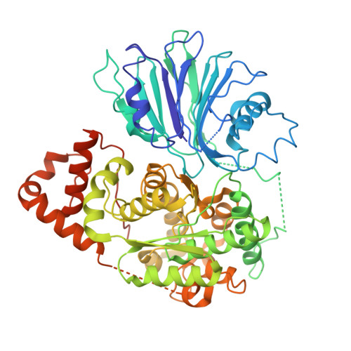
8SUE | pdb_00008sue

Human asparagine synthetase (apo-ASNS)


Experimental Data Snapshot

  • Method: ELECTRON MICROSCOPY
  • Resolution: 3.50 Å
  • Aggregation State: PARTICLE 
  • Reconstruction Method: SINGLE PARTICLE 

Starting Model: experimental
View more details

wwPDB Validation 3D Report Full Report

Validation slider image for 8SUE

This is version 1.1 of the entry. See complete history. 

Literature

3D variability analysis reveals a hidden conformational change controlling ammonia transport in human asparagine synthetase.

Coricello, A., Nardone, A.J., Lupia, A., Gratteri, C., Vos, M., Chaptal, V., Alcaro, S., Zhu, W., Takagi, Y., Richards, N.G.J.

(2024) Nat Commun 15: 10538-10538

  • DOI: https://doi.org/10.1038/s41467-024-54912-9
  • Primary Citation Related Structures: 
    8SUE, 9B6C

  • PubMed Abstract: 

    Advances in X-ray crystallography and cryogenic electron microscopy (cryo-EM) offer the promise of elucidating functionally relevant conformational changes that are not easily studied by other biophysical methods. Here we show that 3D variability analysis (3DVA) of the cryo-EM map for wild-type (WT) human asparagine synthetase (ASNS) identifies a functional role for the Arg-142 side chain and test this hypothesis experimentally by characterizing the R142I variant in which Arg-142 is replaced by isoleucine. Support for Arg-142 playing a role in the intramolecular translocation of ammonia between the active site of the enzyme is provided by the glutamine-dependent synthetase activity of the R142 variant relative to WT ASNS, and MD simulations provide a possible molecular mechanism for these findings. Combining 3DVA with MD simulations is a generally applicable approach to generate testable hypotheses of how conformational changes in buried side chains might regulate function in enzymes.


  • Organizational Affiliation: 
    • Dipartimento di Scienze della Salute, Università "Magna Græcia" di Catanzaro, Catanzaro, Italy.

Macromolecule Content 

  • Total Structure Weight: 128.65 kDa 
  • Atom Count: 8,020 
  • Modeled Residue Count: 1,015 
  • Deposited Residue Count: 1,120 
  • Unique protein chains: 1

Macromolecules

Find similar proteins by:|  3D Structure
Entity ID: 1
MoleculeChains  Sequence LengthOrganismDetailsImage
Asparagine synthetase [glutamine-hydrolyzing]
A, B
560Homo sapiensMutation(s): 0 
Gene Names: ASNS, TS11
EC: 6.3.5.4 (PDB Primary Data), 3.5.1.2 (UniProt)
UniProt & NIH Common Fund Data Resources
Find proteins for P08243 (Homo sapiens)
Explore P08243 
Go to UniProtKB:  P08243
PHAROS:  P08243
GTEx:  ENSG00000070669 
Entity Groups
Sequence Clusters30% Identity50% Identity70% Identity90% Identity95% Identity100% Identity
UniProt GroupP08243
Sequence Annotations
Expand
Reference Sequence

Experimental Data & Validation

Experimental Data

  • Method: ELECTRON MICROSCOPY
  • Resolution: 3.50 Å
  • Aggregation State: PARTICLE 
  • Reconstruction Method: SINGLE PARTICLE 
EM Software:
TaskSoftware PackageVersion
MODEL REFINEMENTPHENIXdev_4893

Structure Validation

View Full Validation Report


Entry History 

& Funding Information

Deposition Data


Funding OrganizationLocationGrant Number
National Institutes of Health/National Institute of General Medical Sciences (NIH/NIGMS)United StatesR01GM111695
National Institutes of Health/National Institute of General Medical Sciences (NIH/NIGMS)United StatesS10OD028723
Centre National de la Recherche Scientifique (CNRS)FranceANR-19-CE11-0023-01
Biotechnology and Biological Sciences Research Council (BBSRC)United KingdomP/0118017/1
European CommissionEuropean UnionFESR FSE 2014- 2020

Revision History  (Full details and data files)

  • Version 1.0: 2024-05-22
    Type: Initial release
  • Version 1.1: 2025-02-05
    Changes: Data collection, Database references, Structure summary