8SSP

AurA bound to danusertib and activating monobody Mb1


Experimental Data Snapshot

  • Method: X-RAY DIFFRACTION
  • Resolution: 2.60 Å
  • R-Value Free: 0.257 
  • R-Value Work: 0.198 
  • R-Value Observed: 0.202 

wwPDB Validation   3D Report Full Report


Ligand Structure Quality Assessment 


This is version 1.0 of the entry. See complete history


Literature

A biophysical framework for double-drugging kinases.

Kim, C.Ludewig, H.Hadzipasic, A.Kutter, S.Nguyen, V.Kern, D.

(2023) Proc Natl Acad Sci U S A 120: e2304611120-e2304611120

  • DOI: https://doi.org/10.1073/pnas.2304611120
  • Primary Citation of Related Structures:  
    8SSN, 8SSO, 8SSP

  • PubMed Abstract: 

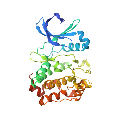
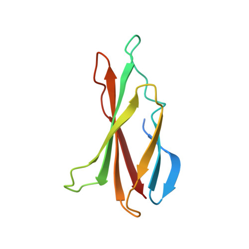
    Selective orthosteric inhibition of kinases has been challenging due to the conserved active site architecture of kinases and emergence of resistance mutants. Simultaneous inhibition of distant orthosteric and allosteric sites, which we refer to as "double-drugging", has recently been shown to be effective in overcoming drug resistance. However, detailed biophysical characterization of the cooperative nature between orthosteric and allosteric modulators has not been undertaken. Here, we provide a quantitative framework for double-drugging of kinases employing isothermal titration calorimetry, Förster resonance energy transfer, coupled-enzyme assays, and X-ray crystallography. We discern positive and negative cooperativity for Aurora A kinase (AurA) and Abelson kinase (Abl) with different combinations of orthosteric and allosteric modulators. We find that a conformational equilibrium shift is the main principle governing cooperativity. Notably, for both kinases, we find a synergistic decrease of the required orthosteric and allosteric drug dosages when used in combination to inhibit kinase activities to clinically relevant inhibition levels. X-ray crystal structures of the double-drugged kinase complexes reveal the molecular principles underlying the cooperative nature of double-drugging AurA and Abl with orthosteric and allosteric inhibitors. Finally, we observe a fully closed conformation of Abl when bound to a pair of positively cooperative orthosteric and allosteric modulators, shedding light on the puzzling abnormality of previously solved closed Abl structures. Collectively, our data provide mechanistic and structural insights into rational design and evaluation of double-drugging strategies.


  • Organizational Affiliation

    Department of Biochemistry, Brandeis University, Waltham, MA 02454.


Macromolecules
Find similar proteins by:  (by identity cutoff)  |  3D Structure
Entity ID: 1
MoleculeChains Sequence LengthOrganismDetailsImage
Aurora kinase A284Homo sapiensMutation(s): 0 
Gene Names: AURKAAIKAIRK1ARK1AURAAYK1BTAKIAK1STK15STK6
EC: 2.7.11.1
UniProt & NIH Common Fund Data Resources
Find proteins for O14965 (Homo sapiens)
Explore O14965 
Go to UniProtKB:  O14965
PHAROS:  O14965
GTEx:  ENSG00000087586 
Entity Groups  
Sequence Clusters30% Identity50% Identity70% Identity90% Identity95% Identity100% Identity
UniProt GroupO14965
Sequence Annotations
Expand
  • Reference Sequence
Find similar proteins by:  (by identity cutoff)  |  3D Structure
Entity ID: 2
MoleculeChains Sequence LengthOrganismDetailsImage
Mb196synthetic constructMutation(s): 0 
Entity Groups  
Sequence Clusters30% Identity50% Identity70% Identity90% Identity95% Identity100% Identity
Sequence Annotations
Expand
  • Reference Sequence
Small Molecules
Ligands 6 Unique
IDChains Name / Formula / InChI Key2D Diagram3D Interactions
627 (Subject of Investigation/LOI)
Query on 627

Download Ideal Coordinates CCD File 
L [auth A]N-[(3E)-5-[(2R)-2-METHOXY-2-PHENYLACETYL]PYRROLO[3,4-C]PYRAZOL-3(5H)-YLIDENE]-4-(4-METHYLPIPERAZIN-1-YL)BENZAMIDE
C26 H30 N6 O3
XKFTZKGMDDZMJI-HSZRJFAPSA-N
MES
Query on MES

Download Ideal Coordinates CCD File 
E [auth A]2-(N-MORPHOLINO)-ETHANESULFONIC ACID
C6 H13 N O4 S
SXGZJKUKBWWHRA-UHFFFAOYSA-N
SO4
Query on SO4

Download Ideal Coordinates CCD File 
D [auth A],
M [auth A]
SULFATE ION
O4 S
QAOWNCQODCNURD-UHFFFAOYSA-L
PO4
Query on PO4

Download Ideal Coordinates CCD File 
H [auth A]PHOSPHATE ION
O4 P
NBIIXXVUZAFLBC-UHFFFAOYSA-K
PDO
Query on PDO

Download Ideal Coordinates CCD File 
I [auth A]1,3-PROPANDIOL
C3 H8 O2
YPFDHNVEDLHUCE-UHFFFAOYSA-N
EDO
Query on EDO

Download Ideal Coordinates CCD File 
C [auth A],
F [auth A],
G [auth A],
J [auth A],
K [auth A]
1,2-ETHANEDIOL
C2 H6 O2
LYCAIKOWRPUZTN-UHFFFAOYSA-N
Experimental Data & Validation

Experimental Data

  • Method: X-RAY DIFFRACTION
  • Resolution: 2.60 Å
  • R-Value Free: 0.257 
  • R-Value Work: 0.198 
  • R-Value Observed: 0.202 
  • Space Group: I 2 2 2
Unit Cell:
Length ( Å )Angle ( ˚ )
a = 75.517α = 90
b = 90.805β = 90
c = 143.13γ = 90
Software Package:
Software NamePurpose
PHENIXrefinement
XDSdata reduction
Aimlessdata scaling
MOLREPphasing

Structure Validation

View Full Validation Report


Ligand Structure Quality Assessment 


Entry History & Funding Information

Deposition Data


Funding OrganizationLocationGrant Number
Howard Hughes Medical Institute (HHMI)United States--

Revision History  (Full details and data files)

  • Version 1.0: 2023-09-06
    Type: Initial release