Structural Analysis and Inhibitor Modeling of Bacterioferritin From Brucella abortus.
Liu, L., Harmon, E.K., Craig, J.K., Yao, H., Battaile, K.P., Johnson, D.K., Subramanian, S., Van Voorhis, W.C., Rivera, M., Lovell, S.(2026) Proteins
- PubMed: 41482512 Search on PubMed
- DOI: https://doi.org/10.1002/prot.70109
- Primary Citation Related Structures:
8SQO, 8SQP, 8SQQ, 8SQR, 8SQT - PubMed Abstract:
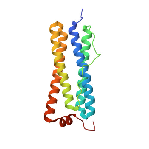
Iron homeostasis in various pathogenic bacteria is regulated by bacterioferritins (Bfr) which function to store Fe 3+ and release Fe 2+ as needed for metabolic processes. The Bfr structure consists of 18 kDa subunits in which dimer pairs bind a heme molecule and are assembled into a highly symmetrical 24-meric spherical structure with an internal core diameter of approximately 80 Å. Release of iron is facilitated by the binding of a 7 kDa [2Fe-2S] ferredoxin (Bfd) to specific sites on the surface of Bfr which transfers electrons to the core thereby reducing the stored Fe 3+ to Fe 2+ for mobilization. The crystal structures of Bfr from Brucella abortus (Ba) in the apo and iron bound forms are presented and compared with those from Acinetobacter baumannii (Ab) and Pseudomonas aeruginosa (Pa). Additionally, models of the Bfr:Bfd complexes for Ba and Ab are provided and compared with the Pa complex. Finally, compounds known to target the Bfr:Bfd interaction in Pa were docked to the Ba and Ab structures which provided insight regarding the potential binding mode and inhibitory mechanism.
- Protein Structure and X-Ray Crystallography Laboratory, Del Shankel Structural Biology Center, University of Kansas, Kansas, USA.
Organizational Affiliation: