8SQ7

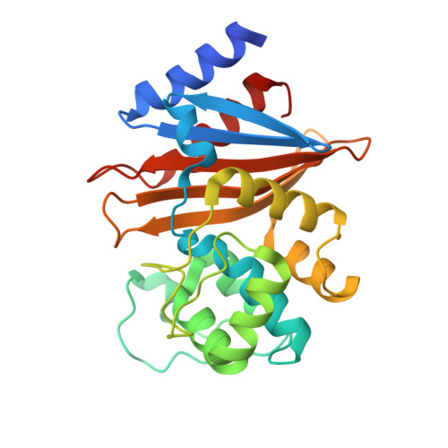
X-ray crystal structure of Acinetobacter baumanii beta-lactamase variant OXA-82 K83D in complex with doripenem


Experimental Data Snapshot

  • Method: X-RAY DIFFRACTION
  • Resolution: 1.78 Å
  • R-Value Free: 0.181 
  • R-Value Work: 0.155 
  • R-Value Observed: 0.156 

wwPDB Validation   3D Report Full Report


Ligand Structure Quality Assessment 


This is version 1.1 of the entry. See complete history


Literature

Structural and Dynamic Features of Acinetobacter baumannii OXA-66 beta-Lactamase Explain Its Stability and Evolution of Novel Variants.

Klamer, Z.L.June, C.M.Wawrzak, Z.Taracila, M.A.Grey, J.A.Benn, A.M.I.Russell, C.P.Bonomo, R.A.Powers, R.A.Leonard, D.A.Szarecka, A.

(2024) J Mol Biol 436: 168603-168603

  • DOI: https://doi.org/10.1016/j.jmb.2024.168603
  • Primary Citation of Related Structures:  
    8SQ7, 8SQ8

  • PubMed Abstract: 

    OXA-66 is a member of the OXA-51 subfamily of class D β-lactamases native to the Acinetobacter genus that includes Acinetobacter baumannii, one of the ESKAPE pathogens and a major cause of drug-resistant nosocomial infections. Although both wild type OXA-66 and OXA-51 have low catalytic activity, they are ubiquitous in the Acinetobacter genomes. OXA-51 is also remarkably thermostable. In addition, newly emerging, single and double amino acid variants show increased activity against carbapenems, indicating that the OXA-51 subfamily is growing and gaining clinical significance. In this study, we used molecular dynamics simulations, X-ray crystallography, and thermal denaturation data to examine and compare the dynamics of OXA-66 wt and its gain-of-function variants: I129L (OXA-83), L167V (OXA-82), P130Q (OXA-109), P130A, and W222L (OXA-234). Our data indicate that OXA-66 wt also has a high melting temperature, and its remarkable stability is due to an extensive and rigid hydrophobic bridge formed by a number of residues around the active site and harbored by the three loops, P, Ω, and β5-β6. Compared to the WT enzyme, the mutants exhibit higher flexibility only in the loop regions, and are more stable than other robust carbapenemases, such as OXA-23 and OXA-24/40. All the mutants show increased rotational flexibility of residues I129 and W222, which allows carbapenems to bind. Overall, our data support the hypothesis that structural features in OXA-51 and OXA-66 promote evolution of multiple highly stable variants with increased clinical relevance in A. baumannii.


  • Organizational Affiliation

    Department of Cell and Molecular Biology, Grand Valley State University, Allendale, MI, USA.


Macromolecules
Find similar proteins by:  (by identity cutoff)  |  3D Structure
Entity ID: 1
MoleculeChains Sequence LengthOrganismDetailsImage
Beta-lactamase OXA-82
A, B, C, D, E
A, B, C, D, E, F, G, H
250Acinetobacter baumanniiMutation(s): 1 
Gene Names: bla-OXA-82blaOXA-82oxa66
UniProt
Find proteins for A8HDA9 (Acinetobacter baumannii)
Explore A8HDA9 
Go to UniProtKB:  A8HDA9
Entity Groups  
Sequence Clusters30% Identity50% Identity70% Identity90% Identity95% Identity100% Identity
UniProt GroupA8HDA9
Sequence Annotations
Expand
  • Reference Sequence
Small Molecules
Ligands 3 Unique
IDChains Name / Formula / InChI Key2D Diagram3D Interactions
4J6 (Subject of Investigation/LOI)
Query on 4J6

Download Ideal Coordinates CCD File 
I [auth A]
K [auth B]
O [auth C]
Q [auth D]
S [auth E]
I [auth A],
K [auth B],
O [auth C],
Q [auth D],
S [auth E],
U [auth F],
W [auth G],
Y [auth H]
(4R,5S)-5-[(2S,3R)-3-hydroxy-1-oxobutan-2-yl]-4-methyl-3-({(3S,5S)-5-[(sulfamoylamino)methyl]pyrrolidin-3-yl}sulfanyl)-4,5-dihydro-1H-pyrrole-2-carboxylic acid
C15 H26 N4 O6 S2
QJKQOMNNBOTMCR-PQTSNVLCSA-N
FLC
Query on FLC

Download Ideal Coordinates CCD File 
J [auth A],
P [auth C],
R [auth D],
T [auth E]
CITRATE ANION
C6 H5 O7
KRKNYBCHXYNGOX-UHFFFAOYSA-K
GOL
Query on GOL

Download Ideal Coordinates CCD File 
AA [auth H]
L [auth B]
M [auth B]
N [auth B]
V [auth F]
AA [auth H],
L [auth B],
M [auth B],
N [auth B],
V [auth F],
X [auth G],
Z [auth H]
GLYCEROL
C3 H8 O3
PEDCQBHIVMGVHV-UHFFFAOYSA-N
Experimental Data & Validation

Experimental Data

  • Method: X-RAY DIFFRACTION
  • Resolution: 1.78 Å
  • R-Value Free: 0.181 
  • R-Value Work: 0.155 
  • R-Value Observed: 0.156 
  • Space Group: P 31
Unit Cell:
Length ( Å )Angle ( ˚ )
a = 70.284α = 90
b = 70.284β = 90
c = 448.355γ = 120
Software Package:
Software NamePurpose
REFMACrefinement
HKL-2000data scaling
HKL-2000data reduction
PHASERphasing

Structure Validation

View Full Validation Report


Ligand Structure Quality Assessment 


Entry History & Funding Information

Deposition Data


Funding OrganizationLocationGrant Number
National Institutes of Health/National Institute Of Allergy and Infectious Diseases (NIH/NIAID)United StatesR01AI072219

Revision History  (Full details and data files)

  • Version 1.0: 2024-05-22
    Type: Initial release
  • Version 1.1: 2024-05-29
    Changes: Database references