8SPI

Crystal structure of chimeric omicron RBD (strain XBB.1.5) complexed with human ACE2


Experimental Data Snapshot

  • Method: X-RAY DIFFRACTION
  • Resolution: 3.06 Å
  • R-Value Free: 0.243 
  • R-Value Work: 0.190 
  • R-Value Observed: 0.193 

wwPDB Validation   3D Report Full Report


Ligand Structure Quality Assessment 


This is version 1.2 of the entry. See complete history


Literature

Structural evolution of SARS-CoV-2 omicron in human receptor recognition.

Zhang, W.Shi, K.Geng, Q.Herbst, M.Wang, M.Huang, L.Bu, F.Liu, B.Aihara, H.Li, F.

(2023) J Virol 97: e0082223-e0082223

  • DOI: https://doi.org/10.1128/jvi.00822-23
  • Primary Citation of Related Structures:  
    8SPH, 8SPI

  • PubMed Abstract: 

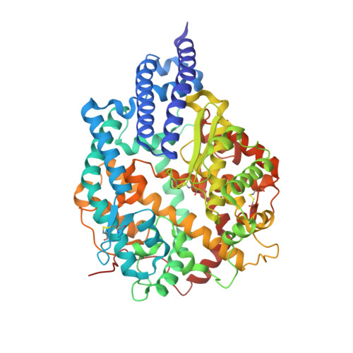
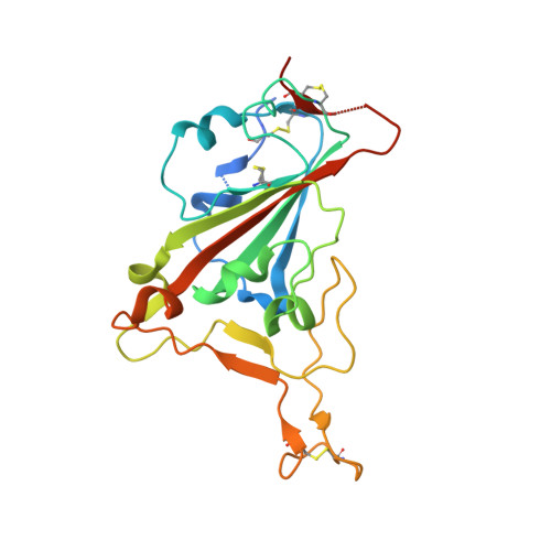
    Understanding the evolutionary strategies of the SARS-CoV-2 omicron variant is crucial for comprehending the COVID-19 pandemic and preventing future coronavirus pandemics. In this study, we determined the crystal structures of the receptor-binding domains (RBDs) from currently circulating omicron subvariants XBB.1 and XBB.1.5 (also the emerging XBB.1.9.1), each complexed with human ACE2. We studied how individual RBD residues evolved structurally in omicron subvariants, specifically how they adapted to human ACE2. Our findings revealed that residues 493 and 496, which exhibited good human ACE2 adaptation in pre-omicron variants, evolved to poor adaptation in early omicron subvariants (but with good adaption to mouse ACE2) and then reverted to good adaptation in recent omicron subvariants. This result is consistent with the hypothesis that non-human animals facilitated the evolution of early omicron subvariants. Additionally, residue 486, which exhibited good human ACE2 adaptation in early omicron subvariants, evolved to poor adaptation in later omicron subvariants and then returned to good adaptation in recent omicron subvariants. This result is consistent with the hypothesis that immune evasion facilitated the evolution of later omicron subvariants. Thus, our study suggests that both non-human animals and immune evasion may have contributed to driving omicron evolution at different stages of the pandemic. IMPORTANCE The sudden emergence and continued evolution of the SARS-CoV-2 omicron variant have left many mysteries unanswered, such as the origin of early omicron subvariants and the factors driving omicron evolution. To address these questions, we studied the crystal structures of human ACE2-bound receptor-binding domains (RBDs) from omicron subvariants XBB.1 and XBB.1.5 (XBB.1.9.1). Our in-depth structural analysis sheds light on how specific RBD mutations adapt to either human or mouse ACE2 and suggests non-human animals and immune evasion may have influenced omicron evolution during different stages of the pandemic. These findings provide valuable insights into the mechanisms underlying omicron evolution, deepen our understanding of the COVID-19 pandemic, and have significant implications for preventing future coronavirus pandemics.


  • Organizational Affiliation

    Department of Pharmacology, University of Minnesota Medical School , Minneapolis, Minnesota, USA.


Macromolecules
Find similar proteins by:  (by identity cutoff)  |  3D Structure
Entity ID: 1
MoleculeChains Sequence LengthOrganismDetailsImage
Angiotensin-converting enzyme 2
A, B
597Homo sapiensMutation(s): 0 
Gene Names: ACE2UNQ868/PRO1885
EC: 3.4.17.23
UniProt & NIH Common Fund Data Resources
Find proteins for Q9BYF1 (Homo sapiens)
Explore Q9BYF1 
Go to UniProtKB:  Q9BYF1
PHAROS:  Q9BYF1
GTEx:  ENSG00000130234 
Entity Groups  
Sequence Clusters30% Identity50% Identity70% Identity90% Identity95% Identity100% Identity
UniProt GroupQ9BYF1
Sequence Annotations
Expand
  • Reference Sequence
Find similar proteins by:  (by identity cutoff)  |  3D Structure
Entity ID: 2
MoleculeChains Sequence LengthOrganismDetailsImage
Spike protein S1C [auth E],
D [auth F]
217Severe acute respiratory syndrome coronavirus 2Mutation(s): 0 
UniProt
Find proteins for P59594 (Severe acute respiratory syndrome coronavirus)
Explore P59594 
Go to UniProtKB:  P59594
Entity Groups  
Sequence Clusters30% Identity50% Identity70% Identity90% Identity95% Identity100% Identity
UniProt GroupP59594
Sequence Annotations
Expand
  • Reference Sequence
Oligosaccharides

Help

Entity ID: 3
MoleculeChains Length2D Diagram Glycosylation3D Interactions
2-acetamido-2-deoxy-beta-D-glucopyranose-(1-4)-2-acetamido-2-deoxy-beta-D-glucopyranoseE [auth C],
H,
I,
K [auth M]
2N-Glycosylation
Glycosylation Resources
GlyTouCan:  G42666HT
GlyCosmos:  G42666HT
GlyGen:  G42666HT
Entity ID: 4
MoleculeChains Length2D Diagram Glycosylation3D Interactions
beta-D-mannopyranose-(1-4)-2-acetamido-2-deoxy-beta-D-glucopyranose-(1-4)-2-acetamido-2-deoxy-beta-D-glucopyranoseF [auth D],
G
3N-Glycosylation
Glycosylation Resources
GlyTouCan:  G15407YE
GlyCosmos:  G15407YE
GlyGen:  G15407YE
Entity ID: 5
MoleculeChains Length2D Diagram Glycosylation3D Interactions
2-acetamido-2-deoxy-beta-D-glucopyranose-(1-3)-beta-D-mannopyranose-(1-4)-2-acetamido-2-deoxy-beta-D-glucopyranose-(1-4)-2-acetamido-2-deoxy-beta-D-glucopyranose
J
4N-Glycosylation
Glycosylation Resources
GlyTouCan:  G25906YW
GlyCosmos:  G25906YW
Small Molecules
Ligands 4 Unique
IDChains Name / Formula / InChI Key2D Diagram3D Interactions
NAG
Query on NAG

Download Ideal Coordinates CCD File 
N [auth A]
O [auth A]
T [auth B]
U [auth B]
V [auth B]
N [auth A],
O [auth A],
T [auth B],
U [auth B],
V [auth B],
W [auth E]
2-acetamido-2-deoxy-beta-D-glucopyranose
C8 H15 N O6
OVRNDRQMDRJTHS-FMDGEEDCSA-N
ZN
Query on ZN

Download Ideal Coordinates CCD File 
L [auth A],
P [auth B]
ZINC ION
Zn
PTFCDOFLOPIGGS-UHFFFAOYSA-N
EDO
Query on EDO

Download Ideal Coordinates CCD File 
R [auth B],
S [auth B]
1,2-ETHANEDIOL
C2 H6 O2
LYCAIKOWRPUZTN-UHFFFAOYSA-N
CL
Query on CL

Download Ideal Coordinates CCD File 
M [auth A],
Q [auth B]
CHLORIDE ION
Cl
VEXZGXHMUGYJMC-UHFFFAOYSA-M
Experimental Data & Validation

Experimental Data

  • Method: X-RAY DIFFRACTION
  • Resolution: 3.06 Å
  • R-Value Free: 0.243 
  • R-Value Work: 0.190 
  • R-Value Observed: 0.193 
  • Space Group: P 1 21 1
Unit Cell:
Length ( Å )Angle ( ˚ )
a = 78.365α = 90
b = 116.205β = 96.6
c = 108.767γ = 90
Software Package:
Software NamePurpose
PHENIXrefinement
autoPROCdata reduction
autoPROCdata scaling
PHASERphasing

Structure Validation

View Full Validation Report


Ligand Structure Quality Assessment 


Entry History & Funding Information

Deposition Data


Funding OrganizationLocationGrant Number
National Institutes of Health/National Institute Of Allergy and Infectious Diseases (NIH/NIAID)United StatesAI089728
National Institutes of Health/National Institute Of Allergy and Infectious Diseases (NIH/NIAID)United StatesAI110700
National Institutes of Health/National Institute of General Medical Sciences (NIH/NIGMS)United StatesGM118047

Revision History  (Full details and data files)

  • Version 1.0: 2023-08-02
    Type: Initial release
  • Version 1.1: 2023-08-30
    Changes: Data collection, Database references
  • Version 1.2: 2023-09-13
    Changes: Database references