Interaction of CYP3A4 with caffeine: First insights into multiple substrate binding.
Sevrioukova, I.F.(2023) J Biological Chem 299: 105117-105117
- PubMed: 37524132 Search on PubMedSearch on PubMed Central
- DOI: https://doi.org/10.1016/j.jbc.2023.105117
- Primary Citation Related Structures:
8SO1, 8SO2 - PubMed Abstract:
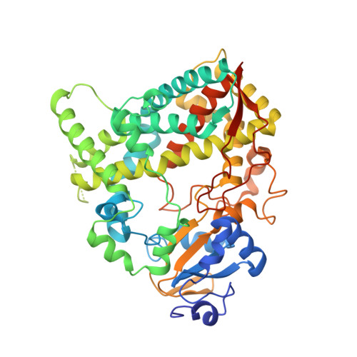
Human cytochrome P450 3A4 (CYP3A4) is a major drug-metabolizing enzyme that shows extreme substrate promiscuity. Moreover, its large and malleable active site can simultaneously accommodate several substrate molecules of the same or different nature, which may lead to cooperative binding and allosteric behavior. Due to difficulty of crystallization of CYP3A4-substrate complexes, it remains unknown how multiple substrates can arrange in the active site. We determined crystal structures of CYP3A4 bound to three and six molecules of caffeine, a psychoactive alkaloid serving as a substrate and modulator of CYP3A4. In the ternary complex, one caffeine binds to the active site suitably for C8-hydroxylation, most preferable for CYP3A4. In the senary complex, three caffeine molecules stack parallel to the heme with the proximal ligand poised for 3-N-demethylation. However, the caffeine stack forms extensive hydrophobic interactions that could preclude product dissociation and multiple turnovers. In both complexes, caffeine is also bound in the substrate channel and on the outer surface known as a peripheral site. At all sites, aromatic stacking with the caffeine ring(s) is likely a dominant interaction, while direct and water-mediated polar contacts provide additional stabilization for the substrate-bound complexes. Protein-ligand interactions via the active site R212, intrachannel T224, and peripheral F219 were experimentally confirmed, and the latter two residues were identified as important for caffeine association. Collectively, the structural, spectral, and mutagenesis data provide valuable insights on the ligand binding mechanism and help better understand how purine-based pharmaceuticals and other aromatic compounds could interact with CYP3A4 and mediate drug-drug interactions.
- Department of Molecular Biology and Biochemistry, University of California, Irvine, California, USA. Electronic address: sevrioui@uci.edu.
Organizational Affiliation: