8SLB

X-ray structure of CorA N-terminal domain in complex with conformation-specific synthetic antibody C12


Experimental Data Snapshot

  • Method: X-RAY DIFFRACTION
  • Resolution: 2.04 Å
  • R-Value Free: 0.218 
  • R-Value Work: 0.166 
  • R-Value Observed: 0.168 

wwPDB Validation   3D Report Full Report


This is version 1.2 of the entry. See complete history


Literature

Conformation-specific Synthetic Antibodies Discriminate Multiple Functional States of the Ion Channel CorA.

Erramilli, S.K.Dominik, P.K.Deneka, D.Tokarz, P.Kim, S.S.Reddy, B.G.Skrobek, B.M.Dalmas, O.Perozo, E.Kossiakoff, A.A.

(2023) J Mol Biol 435: 168192-168192

  • DOI: https://doi.org/10.1016/j.jmb.2023.168192
  • Primary Citation of Related Structures:  
    8SLB

  • PubMed Abstract: 

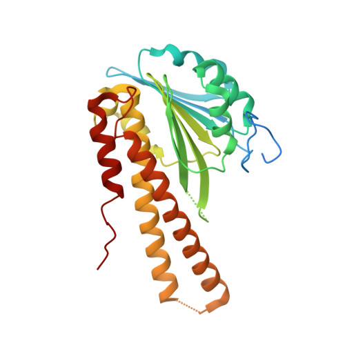
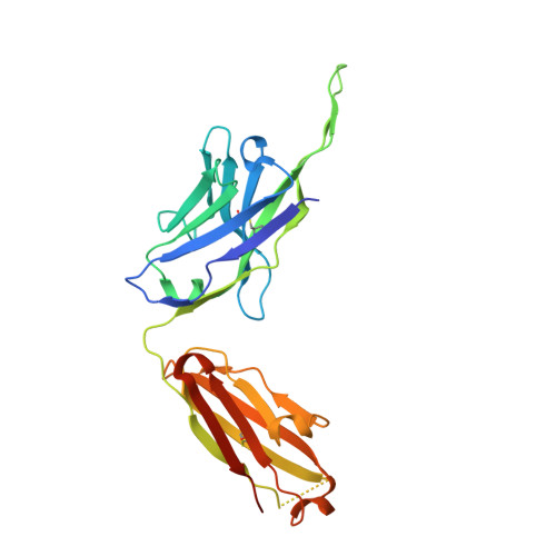
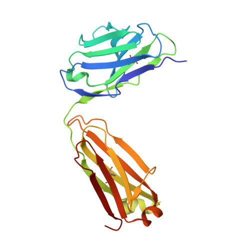
    CorA, the primary magnesium ion channel in prokaryotes and archaea, is a prototypical homopentameric ion channel that undergoes ion-dependent conformational transitions. CorA adopts five-fold symmetric non-conductive states in the presence of high concentrations of Mg 2+ , and highly asymmetric flexible states in its complete absence. However, the latter were of insufficient resolution to be thoroughly characterized. In order to gain additional insights into the relationship between asymmetry and channel activation, we exploited phage display selection strategies to generate conformation-specific synthetic antibodies (sABs) against CorA in the absence of Mg 2+ . Two sABs from these selections, C12 and C18, showed different degrees of Mg 2+ -sensitivity. Through structural, biochemical, and biophysical characterization, we found the sABs are both conformation-specific but probe different features of the channel under open-like conditions. C18 is highly specific to the Mg 2+ -depleted state of CorA and through negative-stain electron microscopy (ns-EM), we show sAB binding reflects the asymmetric arrangement of CorA protomers in Mg 2+ -depleted conditions. We used X-ray crystallography to determine a structure at 2.0 Å resolution of sAB C12 bound to the soluble N-terminal regulatory domain of CorA. The structure shows C12 is a competitive inhibitor of regulatory magnesium binding through its interaction with the divalent cation sensing site. We subsequently exploited this relationship to capture and visualize asymmetric CorA states in different [Mg 2+ ] using ns-EM. We additionally utilized these sABs to provide insights into the energy landscape that governs the ion-dependent conformational transitions of CorA.


  • Organizational Affiliation

    Department of Biochemistry and Molecular Biology, The University of Chicago, Chicago, IL, USA.


Macromolecules
Find similar proteins by:  (by identity cutoff)  |  3D Structure
Entity ID: 1
MoleculeChains Sequence LengthOrganismDetailsImage
Cobalt/magnesium transport protein CorA288Thermotoga maritima MSB8Mutation(s): 0 
Gene Names: corATM_0561
UniProt
Find proteins for Q9WZ31 (Thermotoga maritima (strain ATCC 43589 / DSM 3109 / JCM 10099 / NBRC 100826 / MSB8))
Explore Q9WZ31 
Go to UniProtKB:  Q9WZ31
Entity Groups  
Sequence Clusters30% Identity50% Identity70% Identity90% Identity95% Identity100% Identity
UniProt GroupQ9WZ31
Sequence Annotations
Expand
  • Reference Sequence
Find similar proteins by:  (by identity cutoff)  |  3D Structure
Entity ID: 2
MoleculeChains Sequence LengthOrganismDetailsImage
sAB C12 Heavy ChainB [auth H]240Homo sapiensMutation(s): 0 
Entity Groups  
Sequence Clusters30% Identity50% Identity70% Identity90% Identity95% Identity100% Identity
Sequence Annotations
Expand
  • Reference Sequence
Find similar proteins by:  (by identity cutoff)  |  3D Structure
Entity ID: 3
MoleculeChains Sequence LengthOrganismDetailsImage
sAB C12 Light ChainC [auth L]215Homo sapiensMutation(s): 0 
Entity Groups  
Sequence Clusters30% Identity50% Identity70% Identity90% Identity95% Identity100% Identity
Sequence Annotations
Expand
  • Reference Sequence
Experimental Data & Validation

Experimental Data

  • Method: X-RAY DIFFRACTION
  • Resolution: 2.04 Å
  • R-Value Free: 0.218 
  • R-Value Work: 0.166 
  • R-Value Observed: 0.168 
  • Space Group: I 1 2 1
Unit Cell:
Length ( Å )Angle ( ˚ )
a = 137.338α = 90
b = 44.527β = 108.01
c = 150.634γ = 90
Software Package:
Software NamePurpose
REFMACrefinement
XDSdata reduction
Aimlessdata scaling
PHASERphasing

Structure Validation

View Full Validation Report


Entry History & Funding Information

Deposition Data


Funding OrganizationLocationGrant Number
National Institutes of Health/National Institute of General Medical Sciences (NIH/NIGMS)United StatesGM117372

Revision History  (Full details and data files)

  • Version 1.0: 2023-06-07
    Type: Initial release
  • Version 1.1: 2023-07-12
    Changes: Database references
  • Version 1.2: 2023-07-26
    Changes: Database references