8SK0

Crystal structure of EvdS6 decarboxylase in ligand bound state


Experimental Data Snapshot

  • Method: X-RAY DIFFRACTION
  • Resolution: 1.51 Å
  • R-Value Free: 0.199 
  • R-Value Work: 0.173 
  • R-Value Observed: 0.174 

wwPDB Validation   3D Report Full Report


Ligand Structure Quality Assessment 


This is version 1.0 of the entry. See complete history


Literature

EvdS6 is a bifunctional decarboxylase from the everninomicin gene cluster.

Dulin, C.C.Sharma, P.Frigo, L.Voehler, M.W.Iverson, T.M.Bachmann, B.O.

(2023) J Biol Chem 299: 104893-104893

  • DOI: https://doi.org/10.1016/j.jbc.2023.104893
  • Primary Citation of Related Structures:  
    8SHH, 8SK0

  • PubMed Abstract: 

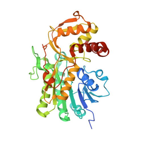
    The everninomicins are bacterially produced antibiotic octasaccharides characterized by the presence of two interglycosidic spirocyclic ortho-δ-lactone (orthoester) moieties. The terminating G- and H-ring sugars, L-lyxose and C-4 branched sugar β-D-eurekanate, are proposed to be biosynthetically derived from nucleotide diphosphate pentose sugar pyranosides; however, the identity of these precursors and their biosynthetic origin remain to be determined. Herein we identify a new glucuronic acid decarboxylase from Micromonospora belonging to the superfamily of short-chain dehydrogenase/reductase enzymes, EvdS6. Biochemical characterization demonstrated that EvdS6 is an NAD + -dependent bifunctional enzyme that produces a mixture of two products, differing in the sugar C-4 oxidation state. This product distribution is atypical for glucuronic acid decarboxylating enzymes, most of which favor production of the reduced sugar and a minority of which favor release of the oxidized product. Spectroscopic and stereochemical analysis of reaction products revealed that the first product released is the oxidatively produced 4-keto-D-xylose and the second product is the reduced D-xylose. X-ray crystallographic analysis of EvdS6 at 1.51 Å resolution with bound co-factor and TDP demonstrated that the overall geometry of the EvdS6 active site is conserved with other SDR enzymes and enabled studies probing structural determinants for the reductive half of the net neutral catalytic cycle. Critical active site threonine and aspartate residues were unambiguously identified as essential in the reductive step of the reaction and resulted in enzyme variants producing almost exclusively the keto sugar. This work defines potential precursors for the G-ring L-lyxose and resolves likely origins of the H-ring β-D-eurekanate sugar precursor.


  • Organizational Affiliation

    Department of Chemistry, Vanderbilt University, Nashville, Tennessee, USA.


Macromolecules
Find similar proteins by:  (by identity cutoff)  |  3D Structure
Entity ID: 1
MoleculeChains Sequence LengthOrganismDetailsImage
dTDP-glucose 4,6-dehydratase
A, B
348Micromonospora carbonaceaMutation(s): 0 
Gene Names: GA0070563_112236
EC: 4.2.1.46
UniProt
Find proteins for A0A1C5ADV9 (Micromonospora carbonacea)
Explore A0A1C5ADV9 
Go to UniProtKB:  A0A1C5ADV9
Entity Groups  
Sequence Clusters30% Identity50% Identity70% Identity90% Identity95% Identity100% Identity
UniProt GroupA0A1C5ADV9
Sequence Annotations
Expand
  • Reference Sequence
Small Molecules
Ligands 5 Unique
IDChains Name / Formula / InChI Key2D Diagram3D Interactions
NAD (Subject of Investigation/LOI)
Query on NAD

Download Ideal Coordinates CCD File 
E [auth A],
H [auth B]
NICOTINAMIDE-ADENINE-DINUCLEOTIDE
C21 H27 N7 O14 P2
BAWFJGJZGIEFAR-NNYOXOHSSA-N
TYD (Subject of Investigation/LOI)
Query on TYD

Download Ideal Coordinates CCD File 
F [auth A]THYMIDINE-5'-DIPHOSPHATE
C10 H16 N2 O11 P2
UJLXYODCHAELLY-XLPZGREQSA-N
FLC (Subject of Investigation/LOI)
Query on FLC

Download Ideal Coordinates CCD File 
I [auth B]CITRATE ANION
C6 H5 O7
KRKNYBCHXYNGOX-UHFFFAOYSA-K
PEG
Query on PEG

Download Ideal Coordinates CCD File 
G [auth B]DI(HYDROXYETHYL)ETHER
C4 H10 O3
MTHSVFCYNBDYFN-UHFFFAOYSA-N
GOL
Query on GOL

Download Ideal Coordinates CCD File 
C [auth A],
D [auth A]
GLYCEROL
C3 H8 O3
PEDCQBHIVMGVHV-UHFFFAOYSA-N
Experimental Data & Validation

Experimental Data

  • Method: X-RAY DIFFRACTION
  • Resolution: 1.51 Å
  • R-Value Free: 0.199 
  • R-Value Work: 0.173 
  • R-Value Observed: 0.174 
  • Space Group: P 21 21 21
Unit Cell:
Length ( Å )Angle ( ˚ )
a = 59.984α = 90
b = 76.405β = 90
c = 148.556γ = 90
Software Package:
Software NamePurpose
PHENIXrefinement
HKL-2000data reduction
HKL-2000data scaling
PHENIXphasing

Structure Validation

View Full Validation Report


Ligand Structure Quality Assessment 


Entry History & Funding Information

Deposition Data


Funding OrganizationLocationGrant Number
National Institutes of Health/National Institute Of Allergy and Infectious Diseases (NIH/NIAID)United States1R01AI140400
American Heart AssociationUnited States19POST34450093

Revision History  (Full details and data files)

  • Version 1.0: 2023-08-09
    Type: Initial release