8SIM

KCNQ1 with voltage sensor in the intermediate conformation


Experimental Data Snapshot

  • Method: ELECTRON MICROSCOPY
  • Resolution: 6.20 Å
  • Aggregation State: PARTICLE 
  • Reconstruction Method: SINGLE PARTICLE 

wwPDB Validation   3D Report Full Report


This is version 1.0 of the entry. See complete history


Literature

The membrane electric field regulates the PIP 2 -binding site to gate the KCNQ1 channel.

Mandala, V.S.MacKinnon, R.

(2023) Proc Natl Acad Sci U S A 120: e2301985120-e2301985120

  • DOI: https://doi.org/10.1073/pnas.2301985120
  • Primary Citation of Related Structures:  
    8SIK, 8SIM, 8SIN

  • PubMed Abstract: 

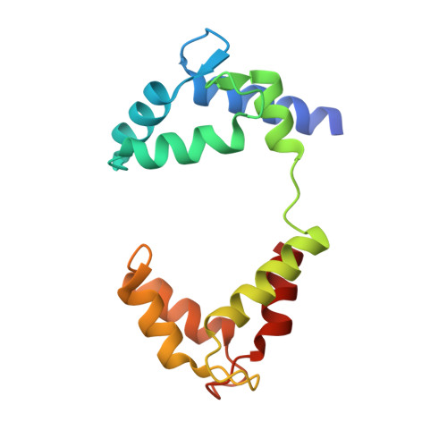
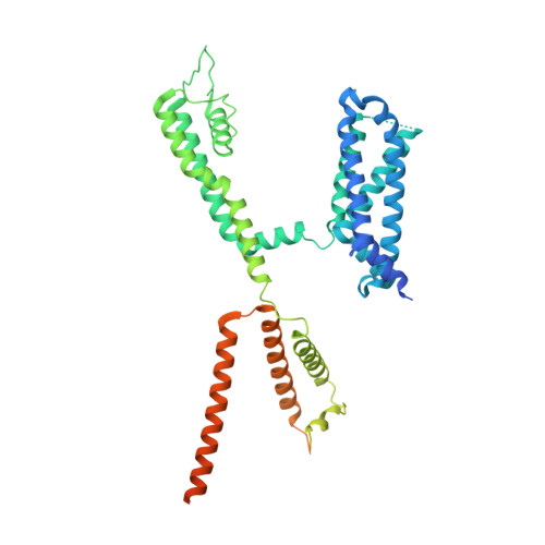
    Voltage-dependent ion channels underlie the propagation of action potentials and other forms of electrical activity in cells. In these proteins, voltage sensor domains (VSDs) regulate opening and closing of the pore through the displacement of their positive-charged S4 helix in response to the membrane voltage. The movement of S4 at hyperpolarizing membrane voltages in some channels is thought to directly clamp the pore shut through the S4-S5 linker helix. The KCNQ1 channel (also known as K v 7.1), which is important for heart rhythm, is regulated not only by membrane voltage but also by the signaling lipid phosphatidylinositol 4,5-bisphosphate (PIP 2 ). KCNQ1 requires PIP 2 to open and to couple the movement of S4 in the VSD to the pore. To understand the mechanism of this voltage regulation, we use cryogenic electron microscopy to visualize the movement of S4 in the human KCNQ1 channel in lipid membrane vesicles with a voltage difference across the membrane, i.e., an applied electric field in the membrane. Hyperpolarizing voltages displace S4 in such a manner as to sterically occlude the PIP 2 -binding site. Thus, in KCNQ1, the voltage sensor acts primarily as a regulator of PIP 2 binding. The voltage sensors' influence on the channel's gate is indirect through the reaction sequence: voltage sensor movement → alter PIP 2 ligand affinity → alter pore opening.


  • Organizational Affiliation

    Laboratory of Molecular Neurobiology and Biophysics, The Rockefeller University, New York, NY 10065.


Macromolecules
Find similar proteins by:  (by identity cutoff)  |  3D Structure
Entity ID: 1
MoleculeChains Sequence LengthOrganismDetailsImage
Calmodulin-1A [auth B],
B [auth D],
C [auth F],
D [auth H]
149Homo sapiensMutation(s): 0 
Gene Names: CALM1CALMCAMCAM1
UniProt & NIH Common Fund Data Resources
Find proteins for P0DP23 (Homo sapiens)
Explore P0DP23 
Go to UniProtKB:  P0DP23
PHAROS:  P0DP23
GTEx:  ENSG00000198668 
Entity Groups  
Sequence Clusters30% Identity50% Identity70% Identity90% Identity95% Identity100% Identity
UniProt GroupP0DP23
Sequence Annotations
Expand
  • Reference Sequence
Find similar proteins by:  (by identity cutoff)  |  3D Structure
Entity ID: 2
MoleculeChains Sequence LengthOrganismDetailsImage
Potassium voltage-gated channel subfamily KQT member 1E,
F [auth A],
G,
H [auth C]
557Homo sapiensMutation(s): 0 
Gene Names: KCNQ1KCNA8KCNA9KVLQT1
Membrane Entity: Yes 
UniProt & NIH Common Fund Data Resources
Find proteins for P51787 (Homo sapiens)
Explore P51787 
Go to UniProtKB:  P51787
PHAROS:  P51787
GTEx:  ENSG00000053918 
Entity Groups  
Sequence Clusters30% Identity50% Identity70% Identity90% Identity95% Identity100% Identity
UniProt GroupP51787
Sequence Annotations
Expand
  • Reference Sequence
Experimental Data & Validation

Experimental Data

  • Method: ELECTRON MICROSCOPY
  • Resolution: 6.20 Å
  • Aggregation State: PARTICLE 
  • Reconstruction Method: SINGLE PARTICLE 

Structure Validation

View Full Validation Report


Entry History & Funding Information

Deposition Data


Funding OrganizationLocationGrant Number
Howard Hughes Medical Institute (HHMI)United States--

Revision History  (Full details and data files)

  • Version 1.0: 2023-05-31
    Type: Initial release