8SH5 | pdb_00008sh5

Crystal structure of 3'cap-independent translation enhancers (CITE) from Pea enation mosaic virus RNA 2 (PEMV2) with Fab BL3-6K170A


Experimental Data Snapshot

  • Method: X-RAY DIFFRACTION
  • Resolution: 2.75 Å
  • R-Value Free: 
    0.297 (Depositor), 0.279 (DCC) 
  • R-Value Work: 
    0.251 (Depositor) 
  • R-Value Observed: 
    0.255 (Depositor) 

Starting Model: experimental
View more details

wwPDB Validation 3D Report Full Report

Validation slider image for 8SH5

This is version 1.3 of the entry. See complete history

Literature

Crystal structure of a cap-independent translation enhancer RNA.

Lewicka, A.Roman, C.Jones, S.Disare, M.Rice, P.A.Piccirilli, J.A.

(2023) Nucleic Acids Res 51: 8891-8907

  • DOI: https://doi.org/10.1093/nar/gkad649
  • Primary Citation Related Structures: 
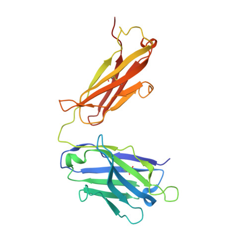
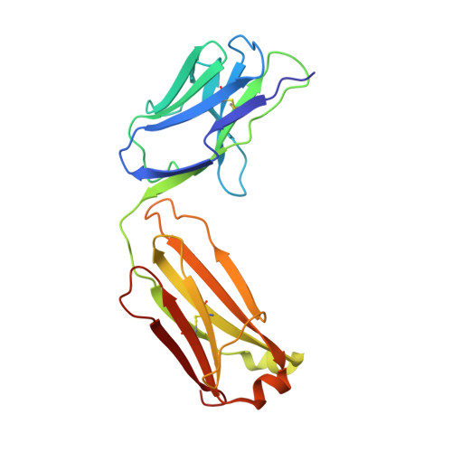
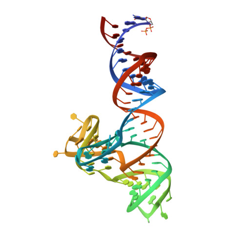
    8SH5

  • PubMed Abstract: 

    In eukaryotic messenger RNAs, the 5' cap structure binds to the translation initiation factor 4E to facilitate early stages of translation. Although many plant viruses lack the 5' cap structure, some contain cap-independent translation elements (CITEs) in their 3' untranslated region. The PTE (Panicum mosaic virus translation element) class of CITEs contains a G-rich asymmetric bulge and a C-rich helical junction that were proposed to interact via formation of a pseudoknot. SHAPE analysis of PTE homologs reveals a highly reactive guanosine residue within the G-rich region proposed to mediate eukaryotic initiation factor 4E (eIF4E) recognition. Here we have obtained the crystal structure of the PTE from Pea enation mosaic virus 2 (PEMV2) RNA in complex with our structural chaperone, Fab BL3-6. The structure reveals that the G-rich and C-rich regions interact through a complex network of interactions distinct from those expected for a pseudoknot. The motif, which contains a short parallel duplex, provides a structural mechanism for how the guanosine is extruded from the core stack to enable eIF4E recognition. Homologous PTE elements harbor a G-rich bulge and a three-way junction and exhibit covariation at crucial positions, suggesting that the PEMV2 tertiary architecture is conserved among these homologs.


  • Organizational Affiliation
    • Department of Biochemistry and Molecular Biology, The University of Chicago, Chicago, IL 60637, USA.

Macromolecule Content 

  • Total Structure Weight: 75.96 kDa 
  • Atom Count: 5,226 
  • Modeled Residue Count: 528 
  • Deposited Residue Count: 531 
  • Unique protein chains: 2
  • Unique nucleic acid chains: 1

Macromolecules


Find similar proteins by:|  3D Structure
Entity ID: 1
MoleculeChains  Sequence LengthOrganismDetailsImage
Fab BL3-6K170A heavy chainA [auth H]228Mus musculusMutation(s): 0 
Find similar proteins by:|  3D Structure
Entity ID: 2
MoleculeChains  Sequence LengthOrganismDetailsImage
Fab BL3-6K170A light chainB [auth L]215Mus musculusMutation(s): 0 
Find similar nucleic acids by:  (by identity cutoff) 
Entity ID: 3
MoleculeChains LengthOrganismImage
RNA (88-MER)C [auth R]88Pea enation mosaic virus 2
Sequence Annotations
Expand
Reference Sequence

Experimental Data & Validation

Experimental Data

  • Method: X-RAY DIFFRACTION
  • Resolution: 2.75 Å
  • R-Value Free:  0.297 (Depositor), 0.279 (DCC) 
  • R-Value Work:  0.251 (Depositor) 
  • R-Value Observed: 0.255 (Depositor) 
Space Group: P 1 21 1
Unit Cell:
Length ( Å )Angle ( ˚ )
a = 63.346α = 90
b = 65.46β = 110.29
c = 89.817γ = 90
Software Package:
Software NamePurpose
PHENIXrefinement
MOSFLMdata reduction
SCALEPACKdata scaling
PHASERphasing

Structure Validation

View Full Validation Report


Entry History 

& Funding Information

Deposition Data


Funding OrganizationLocationGrant Number
National Institutes of Health/National Institute of General Medical Sciences (NIH/NIGMS)United StatesGM102489
National Institutes of Health/National Institute of General Medical Sciences (NIH/NIGMS)United StatesT32GM008720

Revision History  (Full details and data files)

  • Version 1.0: 2023-08-16
    Type: Initial release
  • Version 1.1: 2023-08-30
    Changes: Derived calculations
  • Version 1.2: 2023-09-20
    Changes: Database references
  • Version 1.3: 2024-11-13
    Changes: Structure summary