8SGO | pdb_00008sgo

Human GABAA receptor alpha1-beta2-gamma2 subtype in complex with GABA plus pregnenolone sulfate


Experimental Data Snapshot

  • Method: ELECTRON MICROSCOPY
  • Resolution: 2.65 Å
  • Aggregation State: PARTICLE 
  • Reconstruction Method: SINGLE PARTICLE 

wwPDB Validation 3D Report Full Report

Validation slider image for 8SGO

This is version 2.1 of the entry. See complete history

Literature

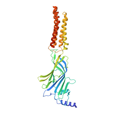
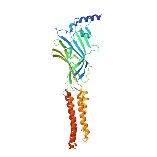
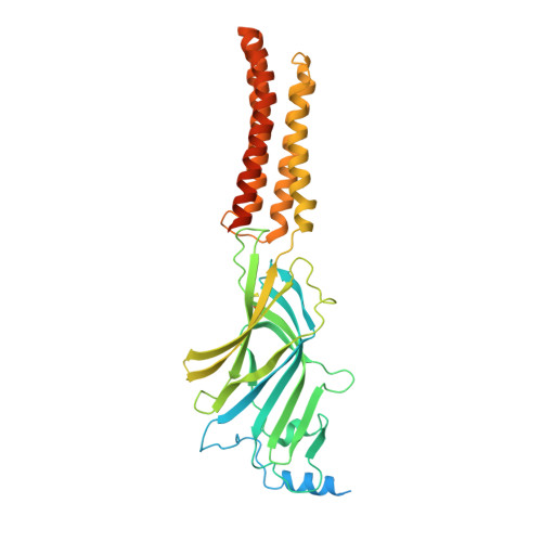
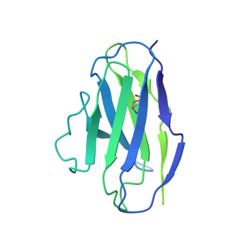
Structural insights into opposing actions of neurosteroids on GABA A receptors.

Legesse, D.H.Fan, C.Teng, J.Zhuang, Y.Howard, R.J.Noviello, C.M.Lindahl, E.Hibbs, R.E.

(2023) Nat Commun 14: 5091-5091

  • DOI: https://doi.org/10.1038/s41467-023-40800-1
  • Primary Citation Related Structures: 
    8SGO, 8SI9, 8SID

  • PubMed Abstract: 

    γ-Aminobutyric acid type A (GABA A ) receptors mediate fast inhibitory signaling in the brain and are targets of numerous drugs and endogenous neurosteroids. A subset of neurosteroids are GABA A receptor positive allosteric modulators; one of these, allopregnanolone, is the only drug approved specifically for treating postpartum depression. There is a consensus emerging from structural, physiological and photolabeling studies as to where positive modulators bind, but how they potentiate GABA activation remains unclear. Other neurosteroids are negative modulators of GABA A receptors, but their binding sites remain debated. Here we present structures of a synaptic GABA A receptor bound to allopregnanolone and two inhibitory sulfated neurosteroids. Allopregnanolone binds at the receptor-bilayer interface, in the consensus potentiator site. In contrast, inhibitory neurosteroids bind in the pore. MD simulations and electrophysiology support a mechanism by which allopregnanolone potentiates channel activity and suggest the dominant mechanism for sulfated neurosteroid inhibition is through pore block.


  • Organizational Affiliation
    • Department of Neuroscience, UT Southwestern Medical Center, Dallas, TX, USA.

Macromolecule Content 

  • Total Structure Weight: 370.17 kDa 
  • Atom Count: 17,742 
  • Modeled Residue Count: 2,121 
  • Deposited Residue Count: 3,195 
  • Unique protein chains: 5

Macromolecules

Find similar proteins by:|  3D Structure
Entity ID: 1
MoleculeChains  Sequence LengthOrganismDetailsImage
Gamma-aminobutyric acid receptor subunit beta-2
A, C
364Homo sapiensMutation(s): 0 
Gene Names: GABRB2
Membrane Entity: Yes 
UniProt & NIH Common Fund Data Resources
Find proteins for P47870 (Homo sapiens)
Explore P47870 
Go to UniProtKB:  P47870
PHAROS:  P47870
GTEx:  ENSG00000145864 
Entity Groups
Sequence Clusters30% Identity50% Identity70% Identity90% Identity95% Identity100% Identity
UniProt GroupP47870
Glycosylation
Glycosylation Sites: 1Go to GlyGen: P47870-2
Sequence Annotations
Expand
Reference Sequence
Find similar proteins by:|  3D Structure
Entity ID: 2
MoleculeChains  Sequence LengthOrganismDetailsImage
Gamma-aminobutyric acid receptor subunit alpha-1
B, D
358Homo sapiensMutation(s): 0 
Gene Names: GABRA1
Membrane Entity: Yes 
UniProt & NIH Common Fund Data Resources
Find proteins for P14867 (Homo sapiens)
Explore P14867 
Go to UniProtKB:  P14867
PHAROS:  P14867
GTEx:  ENSG00000022355 
Entity Groups
Sequence Clusters30% Identity50% Identity70% Identity90% Identity95% Identity100% Identity
UniProt GroupP14867
Glycosylation
Glycosylation Sites: 1Go to GlyGen: P14867-1
Sequence Annotations
Expand
Reference Sequence
Find similar proteins by:|  3D Structure
Entity ID: 3
MoleculeChains  Sequence LengthOrganismDetailsImage
Gamma-aminobutyric acid receptor subunit gamma-2417Homo sapiensMutation(s): 0 
Gene Names: GABRG2
Membrane Entity: Yes 
UniProt & NIH Common Fund Data Resources
Find proteins for P18507 (Homo sapiens)
Explore P18507 
Go to UniProtKB:  P18507
PHAROS:  P18507
GTEx:  ENSG00000113327 
Entity Groups
Sequence Clusters30% Identity50% Identity70% Identity90% Identity95% Identity100% Identity
UniProt GroupP18507
Glycosylation
Glycosylation Sites: 1Go to GlyGen: P18507-2
Sequence Annotations
Expand
Reference Sequence
Find similar proteins by:|  3D Structure
Entity ID: 4
MoleculeChains  Sequence LengthOrganismDetailsImage
Kappa Fab Light ChainF [auth I],
H [auth L]
213Mus musculusMutation(s): 0 
Find similar proteins by:|  3D Structure
Entity ID: 5
MoleculeChains  Sequence LengthOrganismDetailsImage
IgG2b Fab Heavy ChainG [auth J],
I [auth K]
454Mus musculusMutation(s): 0 

Oligosaccharides

Help  
Entity ID: 6
MoleculeChains Length2D Diagram GlycosylationD Interactions
beta-D-mannopyranose-(1-4)-2-acetamido-2-deoxy-beta-D-glucopyranose-(1-4)-2-acetamido-2-deoxy-beta-D-glucopyranoseJ [auth F],
L [auth H],
M
3N-Glycosylation
Glycosylation Resources
GlyTouCan: G15407YE
GlyCosmos: G15407YE
GlyGen: G15407YE
Entity ID: 7
MoleculeChains Length2D Diagram GlycosylationD Interactions
alpha-D-mannopyranose-(1-2)-alpha-D-mannopyranose-(1-2)-alpha-D-mannopyranose-(1-3)-[alpha-D-mannopyranose-(1-2)-alpha-D-mannopyranose-(1-6)-[alpha-D-mannopyranose-(1-3)]alpha-D-mannopyranose-(1-6)]beta-D-mannopyranose-(1-4)-2-acetamido-2-deoxy-beta-D-glucopyranose-(1-4)-2-acetamido-2-deoxy-beta-D-glucopyranoseK [auth G]10N-Glycosylation
Glycosylation Resources
GlyTouCan: G40702WU
GlyCosmos: G40702WU
GlyGen: G40702WU
Entity ID: 8
MoleculeChains Length2D Diagram GlycosylationD Interactions
2-acetamido-2-deoxy-beta-D-glucopyranose-(1-4)-2-acetamido-2-deoxy-beta-D-glucopyranose
N
2N-Glycosylation
Glycosylation Resources
GlyTouCan: G42666HT
GlyCosmos: G42666HT
GlyGen: G42666HT

Small Molecules

Ligands 5 Unique
IDChains Name / Formula / InChI Key2D Diagram3D Interactions
Q3G

Query on Q3G


Download:Ideal Coordinates CCD File
V [auth C]O-[(R)-[(2S)-2-(hexadecanoyloxy)-3-(octadecanoyloxy)propoxy](hydroxy)phosphoryl]-D-serine
C40 H78 N O10 P
WZFUPCSEUKNOBF-PQQNNWGCSA-N
PTY

Query on PTY


Download:Ideal Coordinates CCD File
Q [auth B]
R [auth B]
S [auth B]
W [auth C]
X [auth D]
Q [auth B],
R [auth B],
S [auth B],
W [auth C],
X [auth D],
Y [auth D]
PHOSPHATIDYLETHANOLAMINE
C40 H80 N O8 P
NJGIRBISCGPRPF-KXQOOQHDSA-N
A8W
(Subject of Investigation/LOI)

Query on A8W


Download:Ideal Coordinates CCD File
Z [auth E]Pregnenolone sulfate
C21 H32 O5 S
DIJBBUIOWGGQOP-QGVNFLHTSA-N
NAG

Query on NAG


Download:Ideal Coordinates CCD File
O [auth A],
T [auth C]
2-acetamido-2-deoxy-beta-D-glucopyranose
C8 H15 N O6
OVRNDRQMDRJTHS-FMDGEEDCSA-N
ABU

Query on ABU


Download:Ideal Coordinates CCD File
P [auth A],
U [auth C]
GAMMA-AMINO-BUTANOIC ACID
C4 H9 N O2
BTCSSZJGUNDROE-UHFFFAOYSA-N
Binding Affinity Annotations 
IDSourceBinding Affinity
ABU BindingDB:  8SGO Ki: min: 21.37, max: 77.62 (nM) from 4 assay(s)
IC50: 22 (nM) from 1 assay(s)
EC50: min: 230, max: 8.00e+4 (nM) from 20 assay(s)

Experimental Data & Validation

Experimental Data

  • Method: ELECTRON MICROSCOPY
  • Resolution: 2.65 Å
  • Aggregation State: PARTICLE 
  • Reconstruction Method: SINGLE PARTICLE 
EM Software:
TaskSoftware PackageVersion
MODEL REFINEMENTPHENIX1.19.2_4158
RECONSTRUCTIONRELION3.1

Structure Validation

View Full Validation Report


Entry History 

& Funding Information

Deposition Data


Funding OrganizationLocationGrant Number
National Institutes of Health/National Institute on Drug Abuse (NIH/NIDA)United StatesDA047325
National Institutes of Health/National Institute on Drug Abuse (NIH/NIDA)United StatesDA051176
Welch FoundationUnited StatesI-1812

Revision History  (Full details and data files)

  • Version 1.0: 2023-08-30
    Type: Initial release
  • Version 1.1: 2023-09-06
    Changes: Database references
  • Version 2.0: 2023-11-15
    Changes: Atomic model, Data collection
  • Version 2.1: 2024-11-20
    Changes: Data collection, Structure summary