8SEA

Cryo-EM structure of a double loaded human UBA7-UBE2L6-ISG15 thioester mimetic complex (Form 1)


Experimental Data Snapshot

  • Method: ELECTRON MICROSCOPY
  • Resolution: 3.40 Å
  • Aggregation State: PARTICLE 
  • Reconstruction Method: SINGLE PARTICLE 

wwPDB Validation   3D Report Full Report


This is version 1.0 of the entry. See complete history


Literature

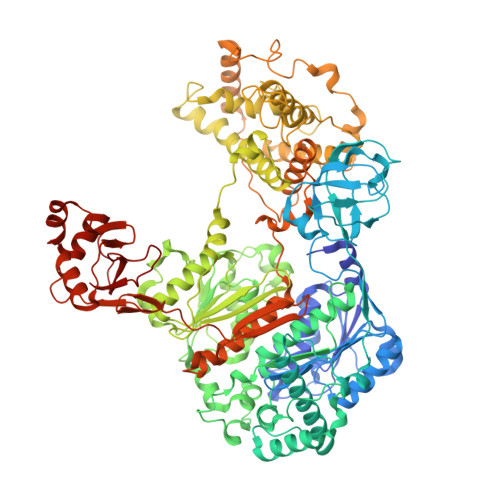
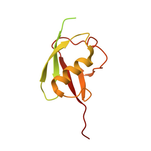
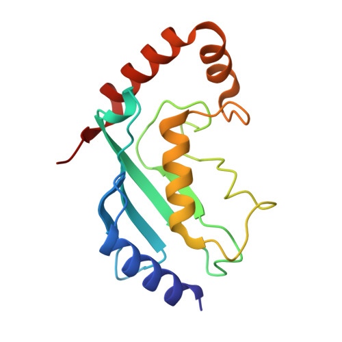
Cryo-EM structures of Uba7 reveal the molecular basis for ISG15 activation and E1-E2 thioester transfer.

Afsar, M.Liu, G.Jia, L.Ruben, E.A.Nayak, D.Sayyad, Z.Bury, P.D.S.Cano, K.E.Nayak, A.Zhao, X.R.Shukla, A.Sung, P.Wasmuth, E.V.Gack, M.U.Olsen, S.K.

(2023) Nat Commun 14: 4786-4786

  • DOI: https://doi.org/10.1038/s41467-023-39780-z
  • Primary Citation of Related Structures:  
    8SE9, 8SEA, 8SEB, 8SV8

  • PubMed Abstract: 

    ISG15 plays a crucial role in the innate immune response and has been well-studied due to its antiviral activity and regulation of signal transduction, apoptosis, and autophagy. ISG15 is a ubiquitin-like protein that is activated by an E1 enzyme (Uba7) and transferred to a cognate E2 enzyme (UBE2L6) to form a UBE2L6-ISG15 intermediate that functions with E3 ligases that catalyze conjugation of ISG15 to target proteins. Despite its biological importance, the molecular basis by which Uba7 catalyzes ISG15 activation and transfer to UBE2L6 is unknown as there is no available structure of Uba7. Here, we present cryo-EM structures of human Uba7 in complex with UBE2L6, ISG15 adenylate, and ISG15 thioester intermediate that are poised for catalysis of Uba7-UBE2L6-ISG15 thioester transfer. Our structures reveal a unique overall architecture of the complex compared to structures from the ubiquitin conjugation pathway, particularly with respect to the location of ISG15 thioester intermediate. Our structures also illuminate the molecular basis for Uba7 activities and for its exquisite specificity for ISG15 and UBE2L6. Altogether, our structural, biochemical, and human cell-based data provide significant insights into the functions of Uba7, UBE2L6, and ISG15 in cells.


  • Organizational Affiliation

    Department of Biochemistry & Structural Biology, University of Texas Health Science Center at San Antonio, San Antonio, TX, 78229, USA.


Macromolecules
Find similar proteins by:  (by identity cutoff)  |  3D Structure
Entity ID: 1
MoleculeChains Sequence LengthOrganismDetailsImage
Ubiquitin-like modifier-activating enzyme 71,012Homo sapiensMutation(s): 0 
Gene Names: UBA7UBE1LUBE2
UniProt & NIH Common Fund Data Resources
Find proteins for P41226 (Homo sapiens)
Explore P41226 
Go to UniProtKB:  P41226
PHAROS:  P41226
GTEx:  ENSG00000182179 
Entity Groups  
Sequence Clusters30% Identity50% Identity70% Identity90% Identity95% Identity100% Identity
UniProt GroupP41226
Sequence Annotations
Expand
  • Reference Sequence
Find similar proteins by:  (by identity cutoff)  |  3D Structure
Entity ID: 2
MoleculeChains Sequence LengthOrganismDetailsImage
Ubiquitin-like protein ISG15
B, D
157Homo sapiensMutation(s): 1 
Gene Names: ISG15G1P2UCRP
UniProt & NIH Common Fund Data Resources
Find proteins for P05161 (Homo sapiens)
Explore P05161 
Go to UniProtKB:  P05161
PHAROS:  P05161
GTEx:  ENSG00000187608 
Entity Groups  
Sequence Clusters30% Identity50% Identity70% Identity90% Identity95% Identity100% Identity
UniProt GroupP05161
Sequence Annotations
Expand
  • Reference Sequence
Find similar proteins by:  (by identity cutoff)  |  3D Structure
Entity ID: 3
MoleculeChains Sequence LengthOrganismDetailsImage
Ubiquitin/ISG15-conjugating enzyme E2 L6152Homo sapiensMutation(s): 3 
Gene Names: UBE2L6UBCH8
EC: 2.3.2.23
UniProt & NIH Common Fund Data Resources
Find proteins for O14933 (Homo sapiens)
Explore O14933 
Go to UniProtKB:  O14933
PHAROS:  O14933
GTEx:  ENSG00000156587 
Entity Groups  
Sequence Clusters30% Identity50% Identity70% Identity90% Identity95% Identity100% Identity
UniProt GroupO14933
Sequence Annotations
Expand
  • Reference Sequence
Small Molecules
Ligands 1 Unique
IDChains Name / Formula / InChI Key2D Diagram3D Interactions
AMP (Subject of Investigation/LOI)
Query on AMP

Download Ideal Coordinates CCD File 
E [auth B]ADENOSINE MONOPHOSPHATE
C10 H14 N5 O7 P
UDMBCSSLTHHNCD-KQYNXXCUSA-N
Experimental Data & Validation

Experimental Data

  • Method: ELECTRON MICROSCOPY
  • Resolution: 3.40 Å
  • Aggregation State: PARTICLE 
  • Reconstruction Method: SINGLE PARTICLE 

Structure Validation

View Full Validation Report


Entry History & Funding Information

Deposition Data


Funding OrganizationLocationGrant Number
National Institutes of Health/National Institute of General Medical Sciences (NIH/NIGMS)United States--

Revision History  (Full details and data files)

  • Version 1.0: 2023-10-11
    Type: Initial release