8S9F

Crystal structure of the kinase domain of Bruton's Tyrosine Kinase bound to dasatinib

  • Classification: TRANSFERASE
  • Organism(s): Mus musculus
  • Expression System: Escherichia coli
  • Mutation(s): Yes 

  • Deposited: 2023-03-28 Released: 2023-08-16 
  • Deposition Author(s): Lin, D.Y., Andreotti, A.H.
  • Funding Organization(s): National Institutes of Health/National Institute Of Allergy and Infectious Diseases (NIH/NIAID)

Experimental Data Snapshot

  • Method: X-RAY DIFFRACTION
  • Resolution: 2.60 Å
  • R-Value Free: 0.284 
  • R-Value Work: 0.242 
  • R-Value Observed: 0.244 

wwPDB Validation   3D Report Full Report


Ligand Structure Quality Assessment 


This is version 1.2 of the entry. See complete history


Literature

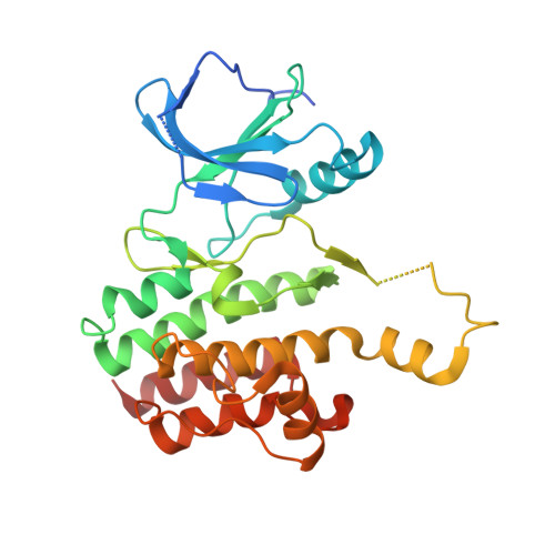
Conformational heterogeneity of the BTK PHTH domain drives multiple regulatory states.

Lin, D.Y.Kueffer, L.E.Juneja, P.Wales, T.E.Engen, J.R.Andreotti, A.H.

(2024) Elife 12

  • DOI: https://doi.org/10.7554/eLife.89489
  • Primary Citation of Related Structures:  
    8GMB, 8S93, 8S9F

  • PubMed Abstract: 

    Full-length Bruton's tyrosine kinase (BTK) has been refractory to structural analysis. The nearest full-length structure of BTK to date consists of the autoinhibited SH3-SH2-kinase core. Precisely how the BTK N-terminal domains (the Pleckstrin homology/Tec homology [PHTH] domain and proline-rich regions [PRR] contain linker) contribute to BTK regulation remains unclear. We have produced crystals of full-length BTK for the first time but despite efforts to stabilize the autoinhibited state, the diffraction data still reveal only the SH3-SH2-kinase core with no electron density visible for the PHTH-PRR segment. Cryo-electron microscopy (cryoEM) data of full-length BTK, on the other hand, provide the first view of the PHTH domain within full-length BTK. CryoEM reconstructions support conformational heterogeneity in the PHTH-PRR region wherein the globular PHTH domain adopts a range of states arrayed around the autoinhibited SH3-SH2-kinase core. On the way to activation, disassembly of the SH3-SH2-kinase core opens a new autoinhibitory site on the kinase domain for PHTH domain binding that is ultimately released upon interaction of PHTH with phosphatidylinositol (3,4,5)-trisphosphate. Membrane-induced dimerization activates BTK and we present here a crystal structure of an activation loop swapped BTK kinase domain dimer that likely represents the conformational state leading to trans-autophosphorylation. Together, these data provide the first structural elucidation of full-length BTK and allow a deeper understanding of allosteric control over the BTK kinase domain during distinct stages of activation.


  • Organizational Affiliation

    Roy J. Carver Department of Biochemistry, Biophysics and Molecular Biology, Iowa State University, Ames, United States.


Macromolecules
Find similar proteins by:  (by identity cutoff)  |  3D Structure
Entity ID: 1
MoleculeChains Sequence LengthOrganismDetailsImage
Tyrosine-protein kinase BTKA [auth B],
B [auth A]
279Mus musculusMutation(s): 3 
Gene Names: BtkBpk
EC: 2.7.10.2
UniProt & NIH Common Fund Data Resources
Find proteins for P35991 (Mus musculus)
Explore P35991 
Go to UniProtKB:  P35991
IMPC:  MGI:88216
Entity Groups  
Sequence Clusters30% Identity50% Identity70% Identity90% Identity95% Identity100% Identity
UniProt GroupP35991
Sequence Annotations
Expand
  • Reference Sequence
Small Molecules
Ligands 1 Unique
IDChains Name / Formula / InChI Key2D Diagram3D Interactions
1N1 (Subject of Investigation/LOI)
Query on 1N1

Download Ideal Coordinates CCD File 
C [auth B],
D [auth A]
N-(2-CHLORO-6-METHYLPHENYL)-2-({6-[4-(2-HYDROXYETHYL)PIPERAZIN-1-YL]-2-METHYLPYRIMIDIN-4-YL}AMINO)-1,3-THIAZOLE-5-CARBOXAMIDE
C22 H26 Cl N7 O2 S
ZBNZXTGUTAYRHI-UHFFFAOYSA-N
Experimental Data & Validation

Experimental Data

  • Method: X-RAY DIFFRACTION
  • Resolution: 2.60 Å
  • R-Value Free: 0.284 
  • R-Value Work: 0.242 
  • R-Value Observed: 0.244 
  • Space Group: P 1 21 1
Unit Cell:
Length ( Å )Angle ( ˚ )
a = 55.237α = 90
b = 110.036β = 99.51
c = 61.142γ = 90
Software Package:
Software NamePurpose
PHENIXrefinement
PHASERphasing
autoPROCdata processing
Cootmodel building
XDSdata scaling
autoPROCdata reduction

Structure Validation

View Full Validation Report


Ligand Structure Quality Assessment 


Entry History & Funding Information

Deposition Data


Funding OrganizationLocationGrant Number
National Institutes of Health/National Institute Of Allergy and Infectious Diseases (NIH/NIAID)United States--

Revision History  (Full details and data files)

  • Version 1.0: 2023-08-16
    Type: Initial release
  • Version 1.1: 2024-01-24
    Changes: Database references
  • Version 1.2: 2024-01-31
    Changes: Database references