8S4P | pdb_00008s4p

Crystal structure of an Ene-reductase from Penicillium steckii


Experimental Data Snapshot

  • Method: X-RAY DIFFRACTION
  • Resolution: 2.29 Å
  • R-Value Free: 
    0.275 (Depositor), 0.267 (DCC) 
  • R-Value Work: 
    0.201 (Depositor), 0.199 (DCC) 
  • R-Value Observed: 
    0.213 (Depositor) 

wwPDB Validation 3D Report Full Report

Validation slider image for 8S4P

Ligand Structure Quality Assessment 


This is version 1.2 of the entry. See complete history

Literature

Recombinant Production and Characterization of Six Ene-reductases from Penicillium steckii.

Damada, P.H.Rozeboom, H.J.Fraaije, M.W.

(2025) Chembiochem 26: e202401007-e202401007

  • DOI: https://doi.org/10.1002/cbic.202401007
  • Primary Citation Related Structures: 
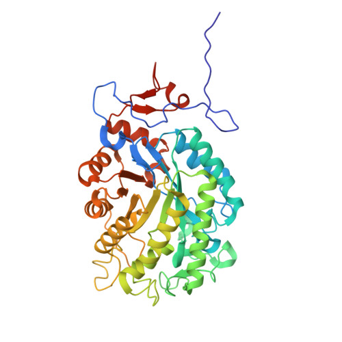
    8S4P

  • PubMed Abstract: 

    Fungi, known for their adaptability, are valuable sources of enzymes, making them promising for biocatalyst discovery. This study explored Penicillium steckii, primarily recognized for secondary meta-bolite production, as a source of ene-reductases (ERs), which reduce α,β-unsaturated compounds. Eleven ER-encoding genes were iden-tified, and plasmids for Escherichia coli expression were generated. Six ERs (PsOYE1-6) were successfully produced and purified as soluble FMN-containing proteins. Sequence analysis classified them into Class II (PsOYE1, PsOYE4, PsOYE6), Class III (PsOYE2, PsOYE3), and Class V (PsOYE5) OYEs. All were active on p-benzo-quinone and maleimide, with varying activity on other substrates. Their pH optima ranged from 6 to 7, and they exhibited moderate thermostability (35-50 °C). PsOYE2 was crystallized, and its 2.3 Å structure revealed a stable dimer with a unique active site. PsOYE3, PsOYE4, and PsOYE5 were tested for R-carvone conversion and stereoselectivity, all favouring one diastereomer. These fungal ERs expand the enzymatic toolbox for biocatalysis, emphasizing the need for tailored strategies based on specific applications.


  • Organizational Affiliation
    • Universidade de Sao Paulo, Laboratório de Química Orgânica e Biocatálise, BRAZIL.

Macromolecule Content 

  • Total Structure Weight: 366.55 kDa 
  • Atom Count: 26,030 
  • Modeled Residue Count: 3,276 
  • Deposited Residue Count: 3,376 
  • Unique protein chains: 1

Macromolecules

Find similar proteins by:|  3D Structure
Entity ID: 1
MoleculeChains  Sequence LengthOrganismDetailsImage
NADH:flavin oxidoreductase/NADH oxidase N-terminal domain-containing protein
A, B, C, D, E
A, B, C, D, E, F, G, H
422Penicillium steckiiMutation(s): 0 
Gene Names: PENSTE_c029G02105
UniProt
Find proteins for A0A1V6SMJ6 (Penicillium steckii)
Explore A0A1V6SMJ6 
Go to UniProtKB:  A0A1V6SMJ6
Entity Groups
Sequence Clusters30% Identity50% Identity70% Identity90% Identity95% Identity100% Identity
UniProt GroupA0A1V6SMJ6
Sequence Annotations
Expand
Reference Sequence

Small Molecules

Ligands 4 Unique
IDChains Name / Formula / InChI Key2D Diagram3D Interactions
FMN

Query on FMN


Download:Ideal Coordinates CCD File
BA [auth H]
I [auth A]
L [auth B]
N [auth C]
P [auth D]
BA [auth H],
I [auth A],
L [auth B],
N [auth C],
P [auth D],
S [auth E],
U [auth F],
Y [auth G]
FLAVIN MONONUCLEOTIDE
C17 H21 N4 O9 P
FVTCRASFADXXNN-SCRDCRAPSA-N
GOL

Query on GOL


Download:Ideal Coordinates CCD File
J [auth A]GLYCEROL
C3 H8 O3
PEDCQBHIVMGVHV-UHFFFAOYSA-N
CA

Query on CA


Download:Ideal Coordinates CCD File
CA [auth H]
O [auth C]
Q [auth D]
V [auth F]
W [auth F]
CA [auth H],
O [auth C],
Q [auth D],
V [auth F],
W [auth F],
Z [auth G]
CALCIUM ION
Ca
BHPQYMZQTOCNFJ-UHFFFAOYSA-N
CL

Query on CL


Download:Ideal Coordinates CCD File
AA [auth G]
DA [auth H]
K [auth A]
M [auth B]
R [auth D]
AA [auth G],
DA [auth H],
K [auth A],
M [auth B],
R [auth D],
T [auth E],
X [auth F]
CHLORIDE ION
Cl
VEXZGXHMUGYJMC-UHFFFAOYSA-M

Experimental Data & Validation

Experimental Data

  • Method: X-RAY DIFFRACTION
  • Resolution: 2.29 Å
  • R-Value Free:  0.275 (Depositor), 0.267 (DCC) 
  • R-Value Work:  0.201 (Depositor), 0.199 (DCC) 
  • R-Value Observed: 0.213 (Depositor) 
Space Group: P 1 21 1
Unit Cell:
Length ( Å )Angle ( ˚ )
a = 107.482α = 90
b = 175.292β = 111.53
c = 107.532γ = 90
Software Package:
Software NamePurpose
PHENIXrefinement
Aimlessdata scaling
XDSdata reduction
PHASERphasing

Structure Validation

View Full Validation Report


Ligand Structure Quality Assessment 


Entry History 

& Funding Information

Deposition Data


Funding OrganizationLocationGrant Number
Not funded--

Revision History  (Full details and data files)

  • Version 1.0: 2025-03-05
    Type: Initial release
  • Version 1.1: 2025-03-19
    Changes: Database references
  • Version 1.2: 2025-04-30
    Changes: Database references