8S2T | pdb_00008s2t

Cryo-EM structure of cell-free synthesized Human beta-1 adrenergic receptor cotranslationally inserted into a lipidic nanodiscs


Experimental Data Snapshot

  • Method: ELECTRON MICROSCOPY
  • Resolution: 3.30 Å
  • Aggregation State: PARTICLE 
  • Reconstruction Method: SINGLE PARTICLE 

Starting Models: experimental, in silico
View more details

wwPDB Validation 3D Report Full Report

Validation slider image for 8S2T

This is version 1.2 of the entry. See complete history

Literature

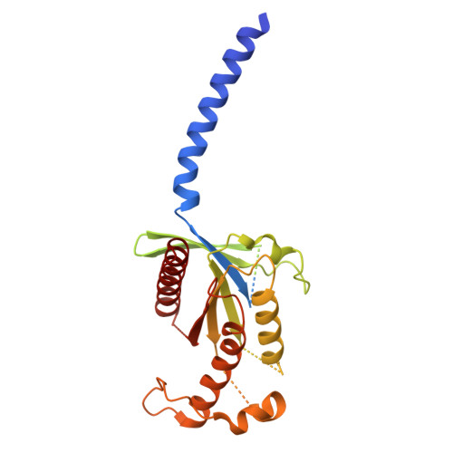
Cryo-EM structure of a cell-free synthesized full-length human beta 1-adrenergic receptor in complex with G s .

Merino, F.Kock, Z.Ermel, U.Dahlhaus, P.Grimm, A.Seybert, A.Kubicek, J.Frangakis, A.S.Dotsch, V.Hilger, D.Bernhard, F.

(2025) Structure 33: 1867

  • DOI: https://doi.org/10.1016/j.str.2025.07.020
  • Primary Citation Related Structures: 
    8S2T

  • PubMed Abstract: 

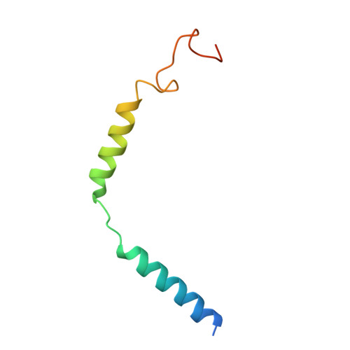
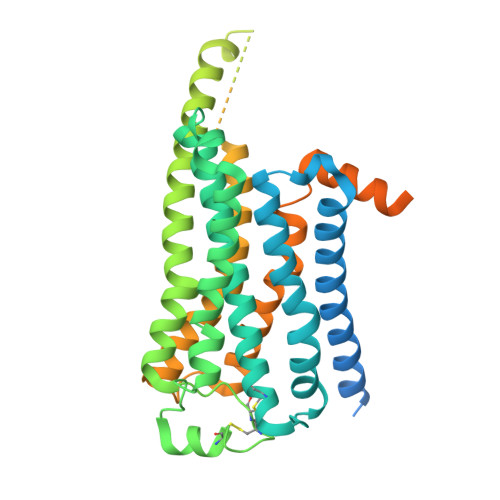
    The third intracellular loop (ICL3) of the β1-adrenergic receptor (β1AR) plays a critical role in regulating G protein coupling, yet the structural basis has remained unclear due to truncations of ICL3 in all available structures of the β1AR in complex with G s or a G protein mimetic nanobody. To address this, we used cell-free cotranslational insertion of full-length human β1AR into nanodiscs and determined its cryo-electron microscopy (cryo-EM) structure in complex with G s . In this structure, ICL3 extends transmembrane helix 5, resulting in enhanced interactions with Gα s and in a slight rotation of the engaged G protein. This repositioning enables new polar interactions between Gα s , ICL2 and helix 8, while ICL1 and helix 8 form additional contacts with Gβ. These structural insights, supported by mutational analysis, demonstrate that ICL3 enhances G protein activation and downstream cAMP signaling by promoting more extensive interactions between the receptor and the heterotrimeric G protein.


  • Organizational Affiliation
    • Cube Biotech GmbH, Monheim, Germany.

Macromolecule Content 

  • Total Structure Weight: 155.67 kDa 
  • Atom Count: 7,955 
  • Modeled Residue Count: 1,002 
  • Deposited Residue Count: 1,404 
  • Unique protein chains: 5

Macromolecules

Find similar proteins by:|  3D Structure
Entity ID: 1
MoleculeChains  Sequence LengthOrganismDetailsImage
Isoform Gnas-2 of Guanine nucleotide-binding protein G(s) subunit alpha isoforms short380Homo sapiensMutation(s): 0 
Gene Names: GNASGNAS1GSP
EC: 3.6.5
UniProt & NIH Common Fund Data Resources
Find proteins for P63092 (Homo sapiens)
Explore P63092 
Go to UniProtKB:  P63092
PHAROS:  P63092
GTEx:  ENSG00000087460 
Entity Groups
Sequence Clusters30% Identity50% Identity70% Identity90% Identity95% Identity100% Identity
UniProt GroupP63092
Sequence Annotations
Expand
Reference Sequence
Find similar proteins by:|  3D Structure
Entity ID: 2
MoleculeChains  Sequence LengthOrganismDetailsImage
Guanine nucleotide-binding protein G(I)/G(S)/G(T) subunit beta-1345Homo sapiensMutation(s): 0 
Gene Names: GNB1
UniProt & NIH Common Fund Data Resources
Find proteins for P62873 (Homo sapiens)
Explore P62873 
Go to UniProtKB:  P62873
PHAROS:  P62873
GTEx:  ENSG00000078369 
Entity Groups
Sequence Clusters30% Identity50% Identity70% Identity90% Identity95% Identity100% Identity
UniProt GroupP62873
Sequence Annotations
Expand
Reference Sequence
Find similar proteins by:|  3D Structure
Entity ID: 3
MoleculeChains  Sequence LengthOrganismDetailsImage
Guanine nucleotide-binding protein G(I)/G(S)/G(O) subunit gamma-268Homo sapiensMutation(s): 0 
Gene Names: GNG2
UniProt & NIH Common Fund Data Resources
Find proteins for P59768 (Homo sapiens)
Explore P59768 
Go to UniProtKB:  P59768
PHAROS:  P59768
GTEx:  ENSG00000186469 
Entity Groups
Sequence Clusters30% Identity50% Identity70% Identity90% Identity95% Identity100% Identity
UniProt GroupP59768
Sequence Annotations
Expand
Reference Sequence
Find similar proteins by:|  3D Structure
Entity ID: 4
MoleculeChains  Sequence LengthOrganismDetailsImage
Nanobody-35D [auth N]134Lama glamaMutation(s): 0 
Find similar proteins by:|  3D Structure
Entity ID: 5
MoleculeChains  Sequence LengthOrganismDetailsImage
Beta-1 adrenergic receptorE [auth R]477Homo sapiensMutation(s): 0 
Gene Names: ADRB1ADRB1RB1AR
UniProt & NIH Common Fund Data Resources
Find proteins for P08588 (Homo sapiens)
Explore P08588 
Go to UniProtKB:  P08588
PHAROS:  P08588
GTEx:  ENSG00000043591 
Entity Groups
Sequence Clusters30% Identity50% Identity70% Identity90% Identity95% Identity100% Identity
UniProt GroupP08588
Sequence Annotations
Expand
Reference Sequence

Small Molecules

Ligands 1 Unique
IDChains Name / Formula / InChI Key2D Diagram3D Interactions
5FW
(Subject of Investigation/LOI)

Query on 5FW


Download:Ideal Coordinates CCD File
F [auth R]ISOPRENALINE
C11 H17 N O3
JWZZKOKVBUJMES-NSHDSACASA-N

Experimental Data & Validation

Experimental Data

  • Method: ELECTRON MICROSCOPY
  • Resolution: 3.30 Å
  • Aggregation State: PARTICLE 
  • Reconstruction Method: SINGLE PARTICLE 
EM Software:
TaskSoftware PackageVersion
MODEL REFINEMENTCoot
MODEL REFINEMENTRosetta
RECONSTRUCTIONRELION

Structure Validation

View Full Validation Report


Entry History 

& Funding Information

Deposition Data


Funding OrganizationLocationGrant Number
German Research Foundation (DFG)GermanyBE1911/9-1
German Research Foundation (DFG)GermanyFR 1653/12-2

Revision History  (Full details and data files)

  • Version 1.0: 2025-05-21
    Type: Initial release
  • Version 1.1: 2025-09-17
    Changes: Data collection, Database references
  • Version 1.2: 2025-11-19
    Changes: Data collection, Database references