8RX4 | pdb_00008rx4

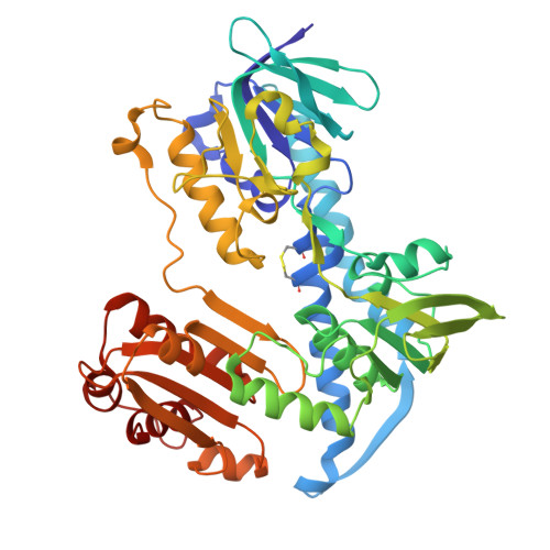
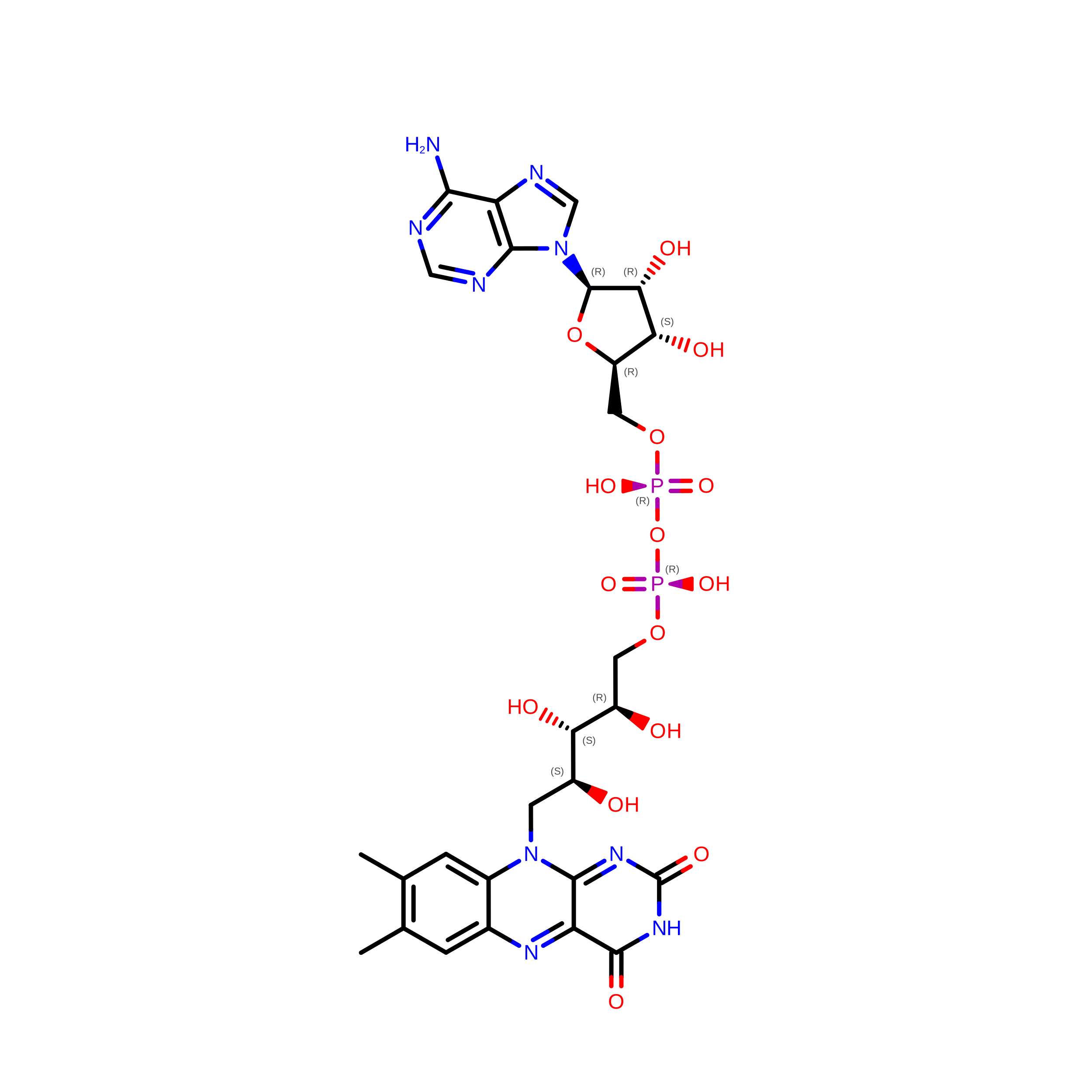
Mycothione reductase from Mycobacterium xenopi in complex with co-factor FAD and redox co-factor NADP(H)


Experimental Data Snapshot

  • Method: X-RAY DIFFRACTION
  • Resolution: 2.34 Å
  • R-Value Free: 
    0.278 (Depositor), 0.277 (DCC) 
  • R-Value Work: 
    0.233 (Depositor), 0.233 (DCC) 
  • R-Value Observed: 
    0.235 (Depositor) 

wwPDB Validation 3D Report Full Report

Validation slider image for 8RX4

Ligand Structure Quality Assessment 


This is version 1.1 of the entry. See complete history. 

Literature

Crystal structure of Mycobacterium tuberculosis mycothione reductase in complex with a specific inhibitor reveals competition with NADP(H) binding.

Oorts, L., Osipov, E.M., Beelen, S., Smiejkowska, N., Van Calster, K., Lamprecht, D., Temmerman, K., Van Aerschot, A., Cappoen, D., Cos, P., Strelkov, S.V.

(2026) FEBS Lett 600: 2143-2153

  • DOI: https://doi.org/10.1002/1873-3468.70336
  • Primary Citation Related Structures: 
    8RX4, 8RX5, 8RX6

  • PubMed Abstract: 

    Tuberculosis (TB) remains a major health threat, while the increasing occurrence of drug-resistant strains underscores the need for new antitubercular drugs. A promising strategy to combat TB is based on disrupting the mycobacterial redox homeostasis by inhibiting an NADPH-dependent oxidoreductase, mycothione reductase (Mtr). Using high-throughput screening, we recently identified potent and selective Mtr inhibitors. Here we report high-resolution X-ray structures of Mtr from Mycobacterium tuberculosis and Mycobacterium xenopi, including the M. tuberculosis enzyme complexed with a novel inhibitor, Respiri-1093. Our findings demonstrate that Respiri-1093 competes with the NADP(H) binding rather than mycothione binding. Analysis of the binding site explains the observed selectivity of the inhibitor towards the M. tuberculosis enzyme. These results provide a structural basis for rational drug design.


  • Organizational Affiliation: 
    • Laboratory of Microbiology, Parasitology and Hygiene, Faculty of Pharmaceutical, Biomedical and Veterinary Sciences, University of Antwerp, Wilrijk, Belgium.

Macromolecule Content 

  • Total Structure Weight: 103.52 kDa 
  • Atom Count: 7,317 
  • Modeled Residue Count: 918 
  • Deposited Residue Count: 918 
  • Unique protein chains: 1

Macromolecules

Find similar proteins by:|  3D Structure
Entity ID: 1
MoleculeChains  Sequence LengthOrganismDetailsImage
Mycothione reductase
A, B
459Mycobacterium xenopiMutation(s): 0 
Gene Names: mtr, I553_7456
EC: 1.8.1.15
UniProt
Find proteins for P9WHH3 (Mycobacterium tuberculosis (strain ATCC 25618 / H37Rv))
Explore P9WHH3 
Go to UniProtKB:  P9WHH3
Entity Groups
Sequence Clusters30% Identity50% Identity70% Identity90% Identity95% Identity100% Identity
UniProt GroupP9WHH3
Sequence Annotations
Expand
Reference Sequence

Experimental Data & Validation

Experimental Data

  • Method: X-RAY DIFFRACTION
  • Resolution: 2.34 Å
  • R-Value Free:  0.278 (Depositor), 0.277 (DCC) 
  • R-Value Work:  0.233 (Depositor), 0.233 (DCC) 
  • R-Value Observed: 0.235 (Depositor) 
Space Group: C 1 2 1
Unit Cell:
Length ( Å )Angle ( ˚ )
a = 128.813α = 90
b = 64.996β = 100.44
c = 137.536γ = 90
Software Package:
Software NamePurpose
PHENIXrefinement
Aimlessdata scaling
XDSdata reduction
PHASERphasing

Structure Validation

View Full Validation Report


Ligand Structure Quality Assessment 


Entry History 

& Funding Information

Deposition Data


Funding OrganizationLocationGrant Number
Research Foundation - Flanders (FWO)BelgiumI007220N
Research Foundation - Flanders (FWO)BelgiumG066619N
European Union (EU)European Union853903

Revision History  (Full details and data files)

  • Version 1.0: 2025-08-20
    Type: Initial release
  • Version 1.1: 2026-10-07
    Changes: Database references