8RUH | pdb_00008ruh

Structure of Oceanobacillus iheyensis group II intron in the presence of K+, Mg2+ and 5'-exon

  • Classification: RNA
  • Organism(s): Oceanobacillus iheyensis
  • Mutation(s): No 

  • Deposited: 2024-01-30 Released: 2024-06-19 
  • Deposition Author(s): Silvestri, I., Marcia, M.
  • Funding Organization(s): Other government, Other private, Fondation ARC, French Infrastructure for Integrated Structural Biology (FRISBI), Grenoble Alliance for Integrated Structural Cell Biology (GRAL), Italian Association for Cancer Research

Experimental Data Snapshot

  • Method: X-RAY DIFFRACTION
  • Resolution: 4.55 Å
  • R-Value Free: 
    0.258 (Depositor), 0.254 (DCC) 
  • R-Value Work: 
    0.184 (Depositor), 0.184 (DCC) 
  • R-Value Observed: 
    0.188 (Depositor) 

Starting Model: experimental
View more details

wwPDB Validation 3D Report Full Report

Validation slider image for 8RUH

This is version 1.1 of the entry. See complete history

Literature

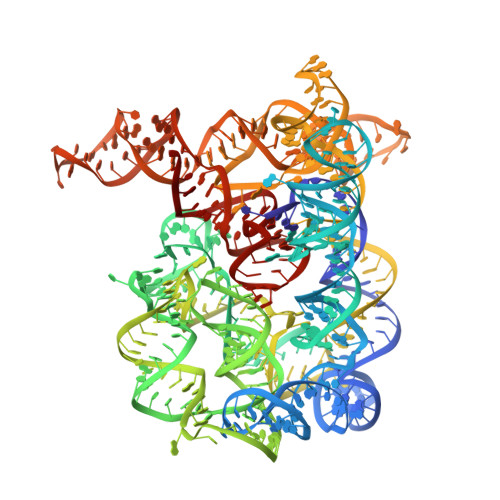
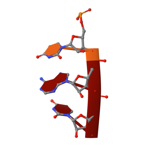
Targeting the conserved active site of splicing machines with specific and selective small molecule modulators.

Silvestri, I.Manigrasso, J.Andreani, A.Brindani, N.Mas, C.Reiser, J.B.Vidossich, P.Martino, G.McCarthy, A.A.De Vivo, M.Marcia, M.

(2024) Nat Commun 15: 4980-4980

  • DOI: https://doi.org/10.1038/s41467-024-48697-0
  • Primary Citation Related Structures: 
    8OLS, 8OLV, 8OLW, 8OLY, 8OLZ, 8OM0, 8RUH, 8RUI, 8RUJ, 8RUK, 8RUL, 8RUM, 8RUN

  • PubMed Abstract: 

    The self-splicing group II introns are bacterial and organellar ancestors of the nuclear spliceosome and retro-transposable elements of pharmacological and biotechnological importance. Integrating enzymatic, crystallographic, and simulation studies, we demonstrate how these introns recognize small molecules through their conserved active site. These RNA-binding small molecules selectively inhibit the two steps of splicing by adopting distinctive poses at different stages of catalysis, and by preventing crucial active site conformational changes that are essential for splicing progression. Our data exemplify the enormous power of RNA binders to mechanistically probe vital cellular pathways. Most importantly, by proving that the evolutionarily-conserved RNA core of splicing machines can recognize small molecules specifically, our work provides a solid basis for the rational design of splicing modulators not only against bacterial and organellar introns, but also against the human spliceosome, which is a validated drug target for the treatment of congenital diseases and cancers.


  • Organizational Affiliation
    • Laboratory of Molecular Modelling & Drug Discovery, Istituto Italiano di Tecnologia, Via Morego 30, 16163, Genoa, Italy.

Macromolecule Content 

  • Total Structure Weight: 130.66 kDa 
  • Atom Count: 8,547 
  • Modeled Residue Count: 393 
  • Deposited Residue Count: 397 
  • Unique nucleic acid chains: 2

Macromolecules

Find similar nucleic acids by:  (by identity cutoff) 
Entity ID: 1
MoleculeChains LengthOrganismImage
Domains 1-5391Oceanobacillus iheyensis
Sequence Annotations
Expand
Reference Sequence
Find similar nucleic acids by:  Sequence
Entity ID: 2
MoleculeChains LengthOrganismImage
5'-exon6Oceanobacillus iheyensis
Sequence Annotations
Expand
Reference Sequence

Small Molecules

Ligands 3 Unique
IDChains Name / Formula / InChI Key2D Diagram3D Interactions
EPE

Query on EPE


Download:Ideal Coordinates CCD File
WA [auth A],
XA [auth A]
4-(2-HYDROXYETHYL)-1-PIPERAZINE ETHANESULFONIC ACID
C8 H18 N2 O4 S
JKMHFZQWWAIEOD-UHFFFAOYSA-N
K

Query on K


Download:Ideal Coordinates CCD File
AA [auth A]
FA [auth A]
GA [auth A]
HA [auth A]
IA [auth A]
AA [auth A],
FA [auth A],
GA [auth A],
HA [auth A],
IA [auth A],
JA [auth A],
KA [auth A],
LA [auth A],
MA [auth A],
NA [auth A],
OA [auth A],
PA [auth A],
QA [auth A],
RA [auth A],
SA [auth A],
TA [auth A],
UA [auth A],
VA [auth A]
POTASSIUM ION
K
NPYPAHLBTDXSSS-UHFFFAOYSA-N
MG

Query on MG


Download:Ideal Coordinates CCD File
BA [auth A]
C [auth A]
CA [auth A]
D [auth A]
DA [auth A]
BA [auth A],
C [auth A],
CA [auth A],
D [auth A],
DA [auth A],
E [auth A],
EA [auth A],
F [auth A],
G [auth A],
H [auth A],
I [auth A],
J [auth A],
K [auth A],
L [auth A],
M [auth A],
N [auth A],
O [auth A],
P [auth A],
Q [auth A],
R [auth A],
S [auth A],
T [auth A],
U [auth A],
V [auth A],
W [auth A],
X [auth A],
Y [auth A],
Z [auth A]
MAGNESIUM ION
Mg
JLVVSXFLKOJNIY-UHFFFAOYSA-N

Experimental Data & Validation

Experimental Data

  • Method: X-RAY DIFFRACTION
  • Resolution: 4.55 Å
  • R-Value Free:  0.258 (Depositor), 0.254 (DCC) 
  • R-Value Work:  0.184 (Depositor), 0.184 (DCC) 
  • R-Value Observed: 0.188 (Depositor) 
Space Group: P 21 21 21
Unit Cell:
Length ( Å )Angle ( ˚ )
a = 90.204α = 90
b = 96.484β = 90
c = 231.693γ = 90
Software Package:
Software NamePurpose
REFMACrefinement
XDSdata reduction
Aimlessdata scaling
PHASERphasing

Structure Validation

View Full Validation Report


Entry History 

& Funding Information

Deposition Data


Funding OrganizationLocationGrant Number
Other governmentITMO Cancer 18CN047-00
Other governmentR21105CC
Other privateFINOVI AAP15
Other governmentCanceropole CLARA Oncostarter
Fondation ARCFrancePJA-20191209284
French Infrastructure for Integrated Structural Biology (FRISBI)FranceANR-10-INSB-05-02
Grenoble Alliance for Integrated Structural Cell Biology (GRAL)FranceANR-10-LABX-49-01
Italian Association for Cancer ResearchItalyIG 23679

Revision History  (Full details and data files)

  • Version 1.0: 2024-06-19
    Type: Initial release
  • Version 1.1: 2024-07-03
    Changes: Database references