8RU3

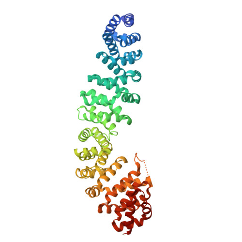
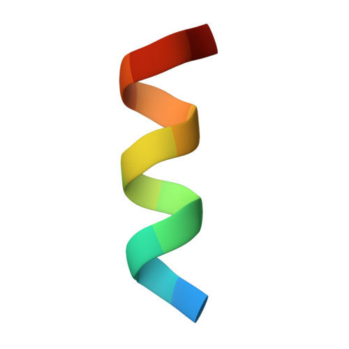
Crystal structure of beta-catenin in complex with alpha-helical peptide inhibitor


Experimental Data Snapshot

  • Method: X-RAY DIFFRACTION
  • Resolution: 2.00 Å
  • R-Value Free: 0.232 
  • R-Value Work: 0.184 

Starting Model: experimental
View more details

wwPDB Validation   3D Report Full Report


This is version 1.2 of the entry. See complete history


Literature

Structure-Based Design of Bicyclic Helical Peptides That Target the Oncogene beta-Catenin.

Yeste-Vazquez, A.Paulussen, F.M.Wendt, M.Klintrot, R.Schulte, C.Wallraven, K.van Gijzel, L.Simeonov, B.van der Gaag, M.Gerber, A.Maric, H.M.Hennig, S.Grossmann, T.N.

(2024) Angew Chem Int Ed Engl 63: e202411749-e202411749

  • DOI: https://doi.org/10.1002/anie.202411749
  • Primary Citation of Related Structures:  
    8RU3, 8RU4

  • PubMed Abstract: 

    The inhibition of intracellular protein-protein interactions is challenging, in particular, when involved interfaces lack pronounced cavities. The transcriptional co-activator protein and oncogene β‑catenin is a prime example of such a challenging target. Despite extensive targeting efforts, available high-affinity binders comprise only large molecular weight Inhibitors. This hampers the further development of therapeutically useful compounds. Herein, we report the design of a considerably smaller peptidomimetic scaffold derived from the α-helical β‑catenin-binding motif of Axin. Sequence maturation and bicyclization provided a stitched peptide with an unprecedented crosslink architecture. The binding mode and site were confirmed by a crystal structure. Further derivatization yielded a β-catenin inhibitor with single-digit micromolar activity in a cell-based assay. This study sheds a light on how to design helix mimetics with reduced molecular weight thereby improving their biological activity.


  • Organizational Affiliation

    VU Amsterdam, Chemisty and Pharmaceutical Sciences, NETHERLANDS.


Macromolecules
Find similar proteins by:  (by identity cutoff)  |  3D Structure
Entity ID: 1
MoleculeChains Sequence LengthOrganismDetailsImage
Catenin beta-1534Homo sapiensMutation(s): 0 
Gene Names: CTNNB1CTNNBOK/SW-cl.35PRO2286
UniProt & NIH Common Fund Data Resources
Find proteins for P35222 (Homo sapiens)
Explore P35222 
Go to UniProtKB:  P35222
PHAROS:  P35222
GTEx:  ENSG00000168036 
Entity Groups  
Sequence Clusters30% Identity50% Identity70% Identity90% Identity95% Identity100% Identity
UniProt GroupP35222
Sequence Annotations
Expand
  • Reference Sequence

Find similar proteins by:  Sequence   |   3D Structure  

Entity ID: 2
MoleculeChains Sequence LengthOrganismDetailsImage
Axin-1B [auth P]14Homo sapiensMutation(s): 4 
UniProt & NIH Common Fund Data Resources
Find proteins for O15169 (Homo sapiens)
Explore O15169 
Go to UniProtKB:  O15169
PHAROS:  O15169
GTEx:  ENSG00000103126 
Entity Groups  
Sequence Clusters30% Identity50% Identity70% Identity90% Identity95% Identity100% Identity
UniProt GroupO15169
Sequence Annotations
Expand
  • Reference Sequence
Experimental Data & Validation

Experimental Data

  • Method: X-RAY DIFFRACTION
  • Resolution: 2.00 Å
  • R-Value Free: 0.232 
  • R-Value Work: 0.184 
  • Space Group: P 21 21 21
Unit Cell:
Length ( Å )Angle ( ˚ )
a = 49.526α = 90
b = 91.255β = 90
c = 112.984γ = 90
Software Package:
Software NamePurpose
REFMACrefinement
Cootmodel building
PHASERphasing
STARANISOdata scaling
xia2.multiplexdata reduction
GDAdata collection

Structure Validation

View Full Validation Report


Entry History & Funding Information

Deposition Data


Funding OrganizationLocationGrant Number
European Research Council (ERC)European Union--

Revision History  (Full details and data files)

  • Version 1.0: 2024-09-04
    Type: Initial release
  • Version 1.1: 2024-10-09
    Changes: Structure summary
  • Version 1.2: 2024-11-20
    Changes: Database references