8RNT | pdb_00008rnt

STRUCTURE OF RIBONUCLEASE T1 COMPLEXED WITH ZINC(II) AT 1.8 ANGSTROMS RESOLUTION: A ZN2+.6H2O.CARBOXYLATE CLATHRATE


Experimental Data Snapshot

  • Method: X-RAY DIFFRACTION
  • Resolution: 1.80 Å
  • R-Value Work: 
    0.136 (DCC) 
  • R-Value Observed: 
    0.140 (Depositor) 

wwPDB Validation 3D Report Full Report

Validation slider image for 8RNT

This is version 1.4 of the entry. See complete history

Literature

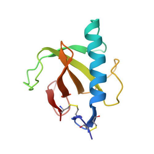
Structure of ribonuclease T1 complexed with zinc(II) at 1.8 A resolution: a Zn2+.6H2O.carboxylate clathrate.

Ding, J.Choe, H.W.Granzin, J.Saenger, W.

(1992) Acta Crystallogr B 48: 185-191

  • DOI: https://doi.org/10.1107/s0108768191013058
  • Primary Citation Related Structures: 
    8RNT

  • PubMed Abstract: 

    In order to study the inhibitory effect of Zn2+ on ribonuclease T1 [RNase T1; Itaya & Inoue (1982). Biochem. J. 207, 357-362], the enzyme was cocrystallized with 2 mM Zn2+, pH 5.2, from a solution containing 55% (v/v) 2-methyl-2,4-pentanediol. The crystals are orthorhombic, P2(1)2(1)2(1), a = 48.71 (1), b = 46.51 (1), c = 41.14 (1) A, Z = 4, V = 93203 A3. The crystal structure was determined by molecular replacement and refined by restrained least-squares methods based on Fhkl for 8291 unique reflections with Fo greater than or equal to 1 sigma (Fo) in the resolution range 10 to 1.8 A and converged at a crystallographic R factor of 0.140. The Zn2+ is not bonded to the active site of RNase T1, probably because the His40 and His92 side chains are protonated. Zn2+ occupies the same site as Ca2+ in a series of crystal structures of free and nucleotide-complexed RNase T1. It is coordinated to Asp15 carboxylate and to six water molecules forming a dodecahedron of square antiprismatic form. The Zn2+...O distances are approximately 2.5 A, suggesting that Zn2+ is clathrated and not coordinated, which would require distances of 2.0 A.


  • Organizational Affiliation
    • Institut für Kristallographie, Freie Universität Berlin, Germany.

Macromolecule Content 

  • Total Structure Weight: 11.16 kDa 
  • Atom Count: 882 
  • Modeled Residue Count: 104 
  • Deposited Residue Count: 104 
  • Unique protein chains: 1

Macromolecules

Find similar proteins by:|  3D Structure
Entity ID: 1
MoleculeChains  Sequence LengthOrganismDetailsImage
RIBONUCLEASE T1104Aspergillus oryzaeMutation(s): 0 
EC: 3.1.27.3 (PDB Primary Data), 4.6.1.24 (UniProt)
UniProt
Find proteins for P00651 (Aspergillus oryzae (strain ATCC 42149 / RIB 40))
Explore P00651 
Go to UniProtKB:  P00651
Entity Groups
Sequence Clusters30% Identity50% Identity70% Identity90% Identity95% Identity100% Identity
UniProt GroupP00651
Sequence Annotations
Expand
Reference Sequence

Small Molecules

Ligands 1 Unique
IDChains Name / Formula / InChI Key2D Diagram3D Interactions
ZN

Query on ZN


Download:Ideal Coordinates CCD File
B [auth A]ZINC ION
Zn
PTFCDOFLOPIGGS-UHFFFAOYSA-N

Experimental Data & Validation

Experimental Data

  • Method: X-RAY DIFFRACTION
  • Resolution: 1.80 Å
  • R-Value Work:  0.136 (DCC) 
  • R-Value Observed: 0.140 (Depositor) 
Space Group: P 21 21 21
Unit Cell:
Length ( Å )Angle ( ˚ )
a = 48.71α = 90
b = 46.51β = 90
c = 41.14γ = 90
Software Package:
Software NamePurpose
PROFFTrefinement

Structure Validation

View Full Validation Report


Entry History 

Deposition Data

Revision History  (Full details and data files)

  • Version 1.0: 1993-01-15
    Type: Initial release
  • Version 1.1: 2008-03-25
    Changes: Version format compliance
  • Version 1.2: 2011-07-13
    Changes: Version format compliance
  • Version 1.3: 2017-11-29
    Changes: Derived calculations, Other
  • Version 1.4: 2024-11-06
    Changes: Data collection, Database references, Derived calculations, Structure summary