8RM0 | pdb_00008rm0

Cryo-EM structure of a Foamy Virus fusion glycoprotein stabilized in the prefusion conformation


Experimental Data Snapshot

  • Method: ELECTRON MICROSCOPY
  • Resolution: 3.80 Å
  • Aggregation State: PARTICLE 
  • Reconstruction Method: SINGLE PARTICLE 

Starting Model: in silico
View more details

wwPDB Validation 3D Report Full Report

Validation slider image for 8RM0

This is version 1.3 of the entry. See complete history

Literature

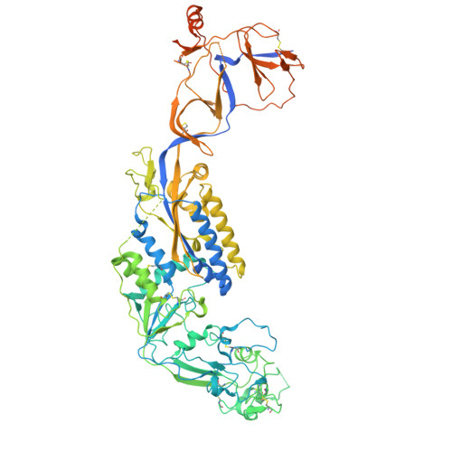
Structures of the Foamy virus fusion protein reveal an unexpected link with the F protein of paramyxo- and pneumoviruses.

Fernandez, I.Bontems, F.Brun, D.Coquin, Y.Goverde, C.A.Correia, B.E.Gessain, A.Buseyne, F.Rey, F.A.Backovic, M.

(2024) Sci Adv 10: eado7035-eado7035

  • DOI: https://doi.org/10.1126/sciadv.ado7035
  • Primary Citation Related Structures: 
    8RM0, 8RM1

  • PubMed Abstract: 

    Foamy viruses (FVs) constitute a subfamily of retroviruses. Their envelope (Env) glycoprotein drives the merger of viral and cellular membranes during entry into cells. The only available structures of retroviral Envs are those from human and simian immunodeficiency viruses from the subfamily of orthoretroviruses, which are only distantly related to the FVs. We report the cryo-electron microscopy structures of the FV Env ectodomain in the pre- and post-fusion states, which unexpectedly demonstrate structural similarity with the fusion protein (F) of paramyxo- and pneumoviruses, implying an evolutionary link between the viral fusogens. We describe the structural features that are unique to the FV Env and propose a mechanistic model for its conformational change, highlighting how the interplay of its structural elements could drive membrane fusion and viral entry. The structural knowledge on the FV Env now provides a framework for functional investigations, which can benefit the design of FV Env variants with improved features for use as gene therapy vectors.


  • Organizational Affiliation
    • Institut Pasteur, Université Paris Cité, CNRS UMR3569, Unité de Virologie Structurale, 75015 Paris, France.

Macromolecule Content 

  • Total Structure Weight: 315.22 kDa 
  • Atom Count: 16,311 
  • Modeled Residue Count: 2,088 
  • Deposited Residue Count: 2,670 
  • Unique protein chains: 1

Macromolecules

Find similar proteins by:|  3D Structure
Entity ID: 1
MoleculeChains  Sequence LengthOrganismDetailsImage
Envelope glycoprotein gp130
A, B, C
890Simian foamy virusMutation(s): 2 
Gene Names: env
UniProt
Find proteins for K7YEW5 (Simian foamy virus)
Explore K7YEW5 
Go to UniProtKB:  K7YEW5
Entity Groups
Sequence Clusters30% Identity50% Identity70% Identity90% Identity95% Identity100% Identity
UniProt GroupK7YEW5
Glycosylation
Glycosylation Sites: 9
Sequence Annotations
Expand
Reference Sequence

Oligosaccharides

Help  
Entity ID: 2
MoleculeChains Length2D Diagram GlycosylationD Interactions
2-acetamido-2-deoxy-beta-D-glucopyranose-(1-4)-2-acetamido-2-deoxy-beta-D-glucopyranose
D, E, G, H, I
D, E, G, H, I, K, L, M, O
2N-Glycosylation
Glycosylation Resources
GlyTouCan: G42666HT
GlyCosmos: G42666HT
GlyGen: G42666HT
Entity ID: 3
MoleculeChains Length2D Diagram GlycosylationD Interactions
alpha-D-mannopyranose-(1-3)-[alpha-D-mannopyranose-(1-6)]beta-D-mannopyranose-(1-4)-2-acetamido-2-deoxy-beta-D-glucopyranose-(1-4)-2-acetamido-2-deoxy-beta-D-glucopyranose
F, J, N
5N-Glycosylation
Glycosylation Resources
GlyTouCan: G22768VO
GlyCosmos: G22768VO
GlyGen: G22768VO

Small Molecules

Ligands 1 Unique
IDChains Name / Formula / InChI Key2D Diagram3D Interactions
NAG

Query on NAG


Download:Ideal Coordinates CCD File
AA [auth C]
BA [auth C]
CA [auth C]
DA [auth C]
P [auth A]
AA [auth C],
BA [auth C],
CA [auth C],
DA [auth C],
P [auth A],
Q [auth A],
R [auth A],
S [auth A],
T [auth A],
U [auth B],
V [auth B],
W [auth B],
X [auth B],
Y [auth B],
Z [auth C]
2-acetamido-2-deoxy-beta-D-glucopyranose
C8 H15 N O6
OVRNDRQMDRJTHS-FMDGEEDCSA-N

Experimental Data & Validation

Experimental Data

  • Method: ELECTRON MICROSCOPY
  • Resolution: 3.80 Å
  • Aggregation State: PARTICLE 
  • Reconstruction Method: SINGLE PARTICLE 
EM Software:
TaskSoftware PackageVersion
MODEL REFINEMENTPHENIX

Structure Validation

View Full Validation Report


Entry History 

& Funding Information

Deposition Data


Funding OrganizationLocationGrant Number
Laboratories of Excellence (LabEx)FranceANR-LBX-62 IBEID

Revision History  (Full details and data files)

  • Version 1.0: 2024-10-23
    Type: Initial release
  • Version 1.1: 2024-10-30
    Changes: Data collection, Database references, Source and taxonomy
  • Version 1.2: 2024-11-20
    Changes: Data collection
  • Version 1.3: 2025-07-02
    Changes: Data collection