A redox switch allows binding of Fe(II) and Fe(III) ions in the cyanobacterial iron-binding protein FutA from Prochlorococcus.
Bolton, R., Machelett, M.M., Stubbs, J., Axford, D., Caramello, N., Catapano, L., Maly, M., Rodrigues, M.J., Cordery, C., Tizzard, G.J., MacMillan, F., Engilberge, S., von Stetten, D., Tosha, T., Sugimoto, H., Worrall, J.A.R., Webb, J.S., Zubkov, M., Coles, S., Mathieu, E., Steiner, R.A., Murshudov, G., Schrader, T.E., Orville, A.M., Royant, A., Evans, G., Hough, M.A., Owen, R.L., Tews, I.(2024) Proc Natl Acad Sci U S A 121: e2308478121-e2308478121
- PubMed: 38489389 Search on PubMedSearch on PubMed Central
- DOI: https://doi.org/10.1073/pnas.2308478121
- Primary Citation Related Structures:
8C4Y, 8OEI, 8OEM, 8OGG, 8RK1 - PubMed Abstract:
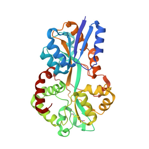
The marine cyanobacterium Prochlorococcus is a main contributor to global photosynthesis, whilst being limited by iron availability. Cyanobacterial genomes generally encode two different types of FutA iron-binding proteins: periplasmic FutA2 ABC transporter subunits bind Fe(III), while cytosolic FutA1 binds Fe(II). Owing to their small size and their economized genome Prochlorococcus ecotypes typically possess a single futA gene. How the encoded FutA protein might bind different Fe oxidation states was previously unknown. Here, we use structural biology techniques at room temperature to probe the dynamic behavior of FutA. Neutron diffraction confirmed four negatively charged tyrosinates, that together with a neutral water molecule coordinate iron in trigonal bipyramidal geometry. Positioning of the positively charged Arg103 side chain in the second coordination shell yields an overall charge-neutral Fe(III) binding state in structures determined by neutron diffraction and serial femtosecond crystallography. Conventional rotation X-ray crystallography using a home source revealed X-ray-induced photoreduction of the iron center with observation of the Fe(II) binding state; here, an additional positioning of the Arg203 side chain in the second coordination shell maintained an overall charge neutral Fe(II) binding site. Dose series using serial synchrotron crystallography and an XFEL X-ray pump-probe approach capture the transition between Fe(III) and Fe(II) states, revealing how Arg203 operates as a switch to accommodate the different iron oxidation states. This switching ability of the Prochlorococcus FutA protein may reflect ecological adaptation by genome streamlining and loss of specialized FutA proteins.
- Biological Sciences, Institute for Life Sciences, University of Southampton, Southampton SO17 1BJ, United Kingdom.
Organizational Affiliation: