8RGY

Serial synchrotron in plate room temperature structure of Dye Type Peroxidase Aa, 8 drops merged


Experimental Data Snapshot

  • Method: X-RAY DIFFRACTION
  • Resolution: 2.07 Å
  • R-Value Free: 0.275 
  • R-Value Work: 0.223 
  • R-Value Observed: 0.226 

wwPDB Validation   3D Report Full Report


Ligand Structure Quality Assessment 


This is version 1.2 of the entry. See complete history


Literature

Efficient in situ screening of and data collection from microcrystals in crystallization plates.

Thompson, A.J.Sanchez-Weatherby, J.Williams, L.J.Mikolajek, H.Sandy, J.Worrall, J.A.R.Hough, M.A.

(2024) Acta Crystallogr D Struct Biol 80: 279-288

  • DOI: https://doi.org/10.1107/S2059798324001955
  • Primary Citation of Related Structures:  
    8RGE, 8RGS, 8RGW, 8RGY

  • PubMed Abstract: 

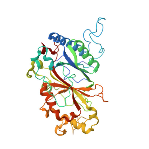
    A considerable bottleneck in serial crystallography at XFEL and synchrotron sources is the efficient production of large quantities of homogenous, well diffracting microcrystals. Efficient high-throughput screening of batch-grown microcrystals and the determination of ground-state structures from different conditions is thus of considerable value in the early stages of a project. Here, a highly sample-efficient methodology to measure serial crystallography data from microcrystals by raster scanning within standard in situ 96-well crystallization plates is described. Structures were determined from very small quantities of microcrystal suspension and the results were compared with those from other sample-delivery methods. The analysis of a two-dimensional batch crystallization screen using this method is also described as a useful guide for further optimization and the selection of appropriate conditions for scaling up microcrystallization.


  • Organizational Affiliation

    Diamond Light Source Ltd, Harwell Science and Innovation Campus, Didcot OX11 0DE, United Kingdom.


Macromolecules
Find similar proteins by:  (by identity cutoff)  |  3D Structure
Entity ID: 1
MoleculeChains Sequence LengthOrganismDetailsImage
Deferrochelatase
A, B
373Streptomyces lividansMutation(s): 0 
Gene Names: SLI_2602
UniProt
Find proteins for A0A7U9DT46 (Streptomyces lividans 1326)
Explore A0A7U9DT46 
Go to UniProtKB:  A0A7U9DT46
Entity Groups  
Sequence Clusters30% Identity50% Identity70% Identity90% Identity95% Identity100% Identity
UniProt GroupA0A7U9DT46
Sequence Annotations
Expand
  • Reference Sequence
Small Molecules
Experimental Data & Validation

Experimental Data

  • Method: X-RAY DIFFRACTION
  • Resolution: 2.07 Å
  • R-Value Free: 0.275 
  • R-Value Work: 0.223 
  • R-Value Observed: 0.226 
  • Space Group: P 1 21 1
Unit Cell:
Length ( Å )Angle ( ˚ )
a = 72.456α = 90
b = 67.759β = 105.69
c = 74.707γ = 90
Software Package:
Software NamePurpose
Cootmodel building
PHENIXrefinement
xia2data reduction
xia2data scaling
PHENIXphasing

Structure Validation

View Full Validation Report


Ligand Structure Quality Assessment 


Entry History & Funding Information

Deposition Data


Funding OrganizationLocationGrant Number
Diamond Light SourceUnited KingdomSTU0436

Revision History  (Full details and data files)

  • Version 1.0: 2023-12-27
    Type: Initial release
  • Version 1.1: 2024-03-27
    Changes: Database references, Structure summary
  • Version 1.2: 2024-04-17
    Changes: Database references