8RE3

Crystal Structure determination of Dye-decolorizing Peroxidase (DyP) mutant M190G from Deinoccoccus radiodurans


Experimental Data Snapshot

  • Method: X-RAY DIFFRACTION
  • Resolution: 1.51 Å
  • R-Value Free: 0.195 
  • R-Value Work: 0.167 
  • R-Value Observed: 0.168 

wwPDB Validation   3D Report Full Report


Ligand Structure Quality Assessment 


This is version 1.0 of the entry. See complete history


Literature

Biochemical, Biophysical, and Structural Analysis of an Unusual DyP from the Extremophile Deinococcus radiodurans.

Frade, K.Silveira, C.M.Salgueiro, B.A.Mendes, S.Martins, L.O.Frazao, C.Todorovic, S.Moe, E.

(2024) Molecules 29

  • DOI: https://doi.org/10.3390/molecules29020358
  • Primary Citation of Related Structures:  
    8RE2, 8RE3

  • PubMed Abstract: 

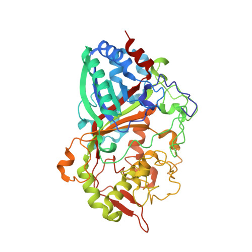
    Dye-decolorizing peroxidases (DyPs) are heme proteins with distinct structural properties and substrate specificities compared to classical peroxidases. Here, we demonstrate that DyP from the extremely radiation-resistant bacterium Deinococcus radiodurans is, like some other homologues, inactive at physiological pH. Resonance Raman (RR) spectroscopy confirms that the heme is in a six-coordinated-low-spin (6cLS) state at pH 7.5 and is thus unable to bind hydrogen peroxide. At pH 4.0, the RR spectra of the enzyme reveal the co-existence of high-spin and low-spin heme states, which corroborates catalytic activity towards H 2 O 2 detected at lower pH. A sequence alignment with other DyPs reveals that Dr DyP possesses a Methionine residue in position five in the highly conserved GXXDG motif. To analyze whether the presence of the Methionine is responsible for the lack of activity at high pH, this residue is substituted with a Glycine. UV-vis and RR spectroscopies reveal that the resulting Dr DyPM190G is also in a 6cLS spin state at pH 7.5, and thus the Methionine does not affect the activity of the protein. The crystal structures of Dr DyP and Dr DyPM190G, determined to 2.20 and 1.53 Å resolution, respectively, nevertheless reveal interesting insights. The high-resolution structure of Dr DyPM190G, obtained at pH 8.5, shows that one hydroxyl group and one water molecule are within hydrogen bonding distance to the heme and the catalytic Asparagine and Arginine. This strong ligand most likely prevents the binding of the H 2 O 2 substrate, reinforcing questions about physiological substrates of this and other DyPs, and about the possible events that can trigger the removal of the hydroxyl group conferring catalytic activity to Dr DyP.


  • Organizational Affiliation

    Instituto de Tecnologia Química e Biológica António Xavier (ITQB-NOVA), Universidade Nova de Lisboa, Av. da Republica (EAN), 2780-157 Oeiras, Portugal.


Macromolecules
Find similar proteins by:  (by identity cutoff)  |  3D Structure
Entity ID: 1
MoleculeChains Sequence LengthOrganismDetailsImage
Peroxidase, putative
A, B
445Deinococcus radioduransMutation(s): 1 
Gene Names: DR_A0145
UniProt
Find proteins for Q9RZ08 (Deinococcus radiodurans (strain ATCC 13939 / DSM 20539 / JCM 16871 / CCUG 27074 / LMG 4051 / NBRC 15346 / NCIMB 9279 / VKM B-1422 / R1))
Explore Q9RZ08 
Go to UniProtKB:  Q9RZ08
Entity Groups  
Sequence Clusters30% Identity50% Identity70% Identity90% Identity95% Identity100% Identity
UniProt GroupQ9RZ08
Sequence Annotations
Expand
  • Reference Sequence
Small Molecules
Ligands 4 Unique
IDChains Name / Formula / InChI Key2D Diagram3D Interactions
HEM (Subject of Investigation/LOI)
Query on HEM

Download Ideal Coordinates CCD File 
C [auth A],
H [auth B]
PROTOPORPHYRIN IX CONTAINING FE
C34 H32 Fe N4 O4
KABFMIBPWCXCRK-RGGAHWMASA-L
CA
Query on CA

Download Ideal Coordinates CCD File 
G [auth A],
L [auth B]
CALCIUM ION
Ca
BHPQYMZQTOCNFJ-UHFFFAOYSA-N
CL
Query on CL

Download Ideal Coordinates CCD File 
F [auth A],
K [auth B]
CHLORIDE ION
Cl
VEXZGXHMUGYJMC-UHFFFAOYSA-M
OH
Query on OH

Download Ideal Coordinates CCD File 
D [auth A],
E [auth A],
I [auth B],
J [auth B]
HYDROXIDE ION
H O
XLYOFNOQVPJJNP-UHFFFAOYSA-M
Experimental Data & Validation

Experimental Data

  • Method: X-RAY DIFFRACTION
  • Resolution: 1.51 Å
  • R-Value Free: 0.195 
  • R-Value Work: 0.167 
  • R-Value Observed: 0.168 
  • Space Group: P 21 21 21
Unit Cell:
Length ( Å )Angle ( ˚ )
a = 82.09α = 90
b = 97.697β = 90
c = 116.037γ = 90
Software Package:
Software NamePurpose
PHENIXrefinement
autoPROCdata reduction
STARANISOdata scaling
PHASERphasing

Structure Validation

View Full Validation Report


Ligand Structure Quality Assessment 


Entry History & Funding Information

Deposition Data


Funding OrganizationLocationGrant Number
Foundation for Science and Technology (FCT)PortugalPTDC/BBBEBB/0122/2014
Foundation for Science and Technology (FCT)PortugalSFRH/BPD/97493/2013
Foundation for Science and Technology (FCT)PortugalCOVID/BD/152598/2022
Foundation for Science and Technology (FCT)PortugalUIDB/04612/2020
Foundation for Science and Technology (FCT)PortugalUIDP/04612/2020
Foundation for Science and Technology (FCT)PortugalLA/P/0087/2020

Revision History  (Full details and data files)

  • Version 1.0: 2024-01-31
    Type: Initial release