8RBF | pdb_00008rbf

CryoEM structure of the post-powerstroke actomyosin-5a complex


Experimental Data Snapshot

  • Method: ELECTRON MICROSCOPY
  • Resolution: 4.20 Å
  • Aggregation State: FILAMENT 
  • Reconstruction Method: SINGLE PARTICLE 

wwPDB Validation   3D Report Full Report


This is version 1.2 of the entry. See complete history


Literature

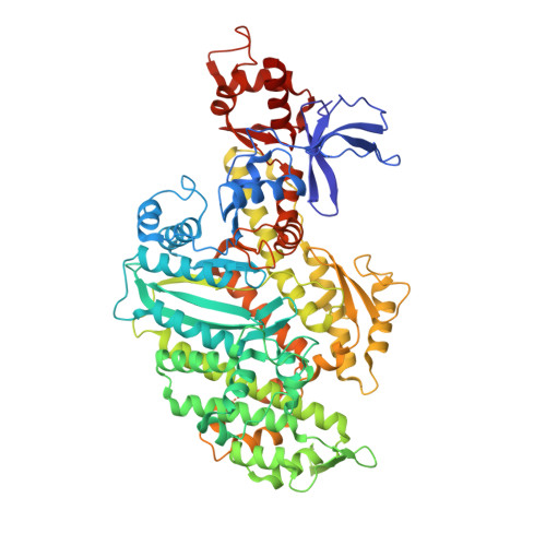
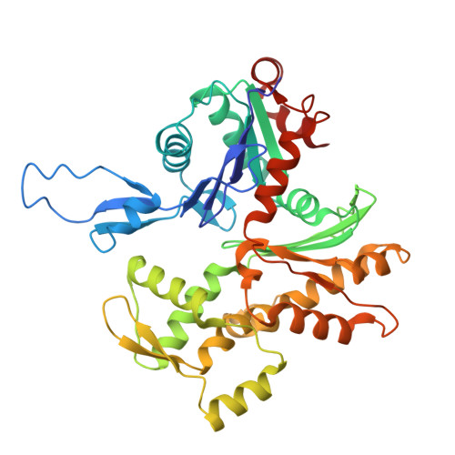
Swinging lever mechanism of myosin directly shown by time-resolved cryo-EM.

Klebl, D.P.McMillan, S.N.Risi, C.Forgacs, E.Virok, B.Atherton, J.L.Harris, S.A.Stofella, M.Winkelmann, D.A.Sobott, F.Galkin, V.E.Knight, P.J.Muench, S.P.Scarff, C.A.White, H.D.

(2025) Nature 642: 519-526

  • DOI: https://doi.org/10.1038/s41586-025-08876-5
  • Primary Citation of Related Structures:  
    8R9V, 8RBF, 8RBG

  • PubMed Abstract: 

    Myosins produce force and movement in cells through interactions with F-actin 1 . Generation of movement is thought to arise through actin-catalysed conversion of myosin from an ATP-generated primed (pre-powerstroke) state to a post-powerstroke state, accompanied by myosin lever swing 2,3 . However, the initial, primed actomyosin state has never been observed, and the mechanism by which actin catalyses myosin ATPase activity is unclear. Here, to address these issues, we performed time-resolved cryogenic electron microscopy (cryo-EM) 4 of a myosin-5 mutant having slow hydrolysis product release 5,6 . Primed actomyosin was predominantly captured 10 ms after mixing primed myosin with F-actin, whereas post-powerstroke actomyosin predominated at 120 ms, with no abundant intermediate states detected. For detailed interpretation, cryo-EM maps were fitted with pseudo-atomic models. Small but critical changes accompany the primed motor binding to actin through its lower 50-kDa subdomain, with the actin-binding cleft open and phosphate release prohibited. Amino-terminal actin interactions with myosin promote rotation of the upper 50-kDa subdomain, closing the actin-binding cleft, and enabling phosphate release. The formation of interactions between the upper 50-kDa subdomain and actin creates the strong-binding interface needed for effective force production. The myosin-5 lever swings through 93°, predominantly along the actin axis, with little twisting. The magnitude of lever swing matches the typical step length of myosin-5 along actin 7 . These time-resolved structures demonstrate the swinging lever mechanism, elucidate structural transitions of the power stroke, and resolve decades of conjecture on how myosins generate movement.


  • Organizational Affiliation
    • School of Biomedical Sciences, Faculty of Biological Sciences, University of Leeds, Leeds, UK.

Macromolecules
Find similar proteins by:  (by identity cutoff)  |  3D Structure
Entity ID: 1
MoleculeChains Sequence LengthOrganismDetailsImage
Unconventional myosin-Va759Mus musculusMutation(s): 1 
Gene Names: Myo5aDilute
UniProt & NIH Common Fund Data Resources
Find proteins for Q99104 (Mus musculus)
Explore Q99104 
Go to UniProtKB:  Q99104
IMPC:  MGI:105976
Entity Groups  
Sequence Clusters30% Identity50% Identity70% Identity90% Identity95% Identity100% Identity
UniProt GroupQ99104
Sequence Annotations
Expand
  • Reference Sequence
Find similar proteins by:  (by identity cutoff)  |  3D Structure
Entity ID: 2
MoleculeChains Sequence LengthOrganismDetailsImage
Actin, alpha skeletal muscle
B, C, D
376Oryctolagus cuniculusMutation(s): 1 
EC: 3.6.4
UniProt
Find proteins for P68135 (Oryctolagus cuniculus)
Explore P68135 
Go to UniProtKB:  P68135
Entity Groups  
Sequence Clusters30% Identity50% Identity70% Identity90% Identity95% Identity100% Identity
UniProt GroupP68135
Sequence Annotations
Expand
  • Reference Sequence
Small Molecules
Ligands 2 Unique
IDChains Name / Formula / InChI Key2D Diagram3D Interactions
ADP (Subject of Investigation/LOI)
Query on ADP

Download Ideal Coordinates CCD File 
F [auth A],
G [auth B],
I [auth C],
K [auth D]
ADENOSINE-5'-DIPHOSPHATE
C10 H15 N5 O10 P2
XTWYTFMLZFPYCI-KQYNXXCUSA-N
MG (Subject of Investigation/LOI)
Query on MG

Download Ideal Coordinates CCD File 
E [auth A],
H [auth B],
J [auth C],
L [auth D]
MAGNESIUM ION
Mg
JLVVSXFLKOJNIY-UHFFFAOYSA-N
Experimental Data & Validation

Experimental Data

  • Method: ELECTRON MICROSCOPY
  • Resolution: 4.20 Å
  • Aggregation State: FILAMENT 
  • Reconstruction Method: SINGLE PARTICLE 

Structure Validation

View Full Validation Report


Entry History & Funding Information

Deposition Data


Funding OrganizationLocationGrant Number
Wellcome TrustUnited Kingdom--
Biotechnology and Biological Sciences Research Council (BBSRC)United Kingdom--
American Heart AssociationUnited States--
Medical Research Council (MRC, United Kingdom)United Kingdom--
British Heart FoundationUnited Kingdom--

Revision History  (Full details and data files)

  • Version 1.0: 2024-12-11
    Type: Initial release
  • Version 1.1: 2025-04-23
    Changes: Data collection, Database references
  • Version 1.2: 2025-06-25
    Changes: Data collection, Database references