8R0T | pdb_00008r0t

STRUCTURE OF THE MOUSE FCGBP DIMER PROTEIN IN ITS SEMIEXTENDED CONFORMATION


Experimental Data Snapshot

  • Method: ELECTRON MICROSCOPY
  • Resolution: 3.90 Å
  • Aggregation State: PARTICLE 
  • Reconstruction Method: SINGLE PARTICLE 

wwPDB Validation   3D Report Full Report


This is version 1.2 of the entry. See complete history


Literature

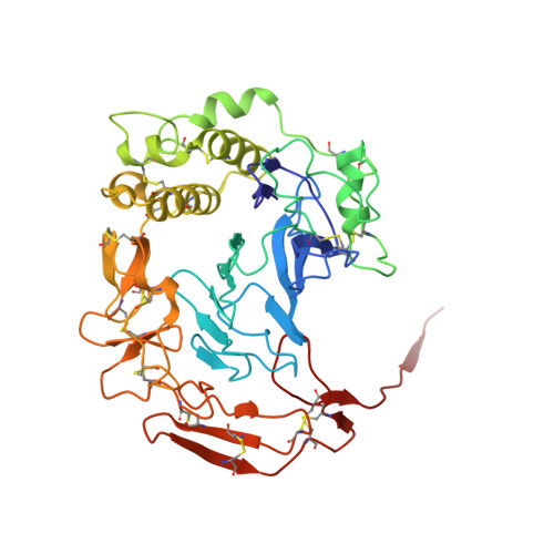
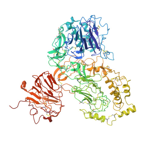
The structure of FCGBP is formed as a disulfide-mediated homodimer between its C-terminal domains.

Ehrencrona, E.Gallego, P.Trillo-Muyo, S.Garcia-Bonete, M.J.Recktenwald, C.V.Hansson, G.C.Johansson, M.E.V.

(2025) FEBS J 292: 582-601

  • DOI: https://doi.org/10.1111/febs.17383
  • Primary Citation of Related Structures:  
    8R0T, 8RDE

  • PubMed Abstract: 

    Mucus in the colon is crucial for intestinal homeostasis by forming a barrier that separates microbes from the epithelium. This is achieved by the structural arrangement of the major mucus proteins, such as MUC2 and FCGBP, both of which are comprised of several von Willebrand D domains (vWD) and assemblies. Numerous disulfide bonds stabilise these domains, and intermolecular bonds generate multimers of MUC2. The oligomeric nature of FCGBP is not known. Human hFCGBP contains 13 vWD domains whereas mouse mFCGBP consists of only 7. We found unpaired cysteines in the vWD1 (human and mouse) and vWD5 (mouse)/vWD11 (human) assemblies which were not involved in disulfide bonds. However, the most C-terminal vWD domains, vWD7 (mouse)/vWD13 (human), formed disulfide-linked dimers. The intermolecular bond between C 5284 and C 5403 of human hFCGBP was observed by using mass spectrometry to generate the dimer. Cryo-EM structure analysis of recombinant mouse mFCGBP revealed a compact dimer with two symmetric intermolecular disulfide bonds between C 2462 and C 2581 , corresponding to the dimerising cysteines in the human hFCGBP. This compact conformation involves interactions between the vWD assemblies, but although the domains involved at the interface are the same, the nature of the interactions differ. Mouse mFCGBP was also found to exist in a semi-extended conformation. These different interactions offer insights into the dynamic nature of the FCGBP homodimer.


  • Organizational Affiliation
    • Department of Medical Biochemistry and Cell Biology, Institute of Biomedicine, University of Gothenburg, Sweden.

Macromolecules
Find similar proteins by:  (by identity cutoff)  |  3D Structure
Entity ID: 1
MoleculeChains Sequence LengthOrganismDetailsImage
Fc fragment of IgG binding protein1,232Mus musculusMutation(s): 0 
Gene Names: Fcgbp
UniProt & NIH Common Fund Data Resources
Find proteins for E9Q0B5 (Mus musculus)
Explore E9Q0B5 
Go to UniProtKB:  E9Q0B5
IMPC:  MGI:2444336
Entity Groups  
Sequence Clusters30% Identity50% Identity70% Identity90% Identity95% Identity100% Identity
UniProt GroupE9Q0B5
Glycosylation
Glycosylation Sites: 1
Sequence Annotations
Expand
  • Reference Sequence
Find similar proteins by:  (by identity cutoff)  |  3D Structure
Entity ID: 2
MoleculeChains Sequence LengthOrganismDetailsImage
Fc fragment of IgG binding protein399Mus musculusMutation(s): 0 
Gene Names: Fcgbp
UniProt & NIH Common Fund Data Resources
Find proteins for E9Q0B5 (Mus musculus)
Explore E9Q0B5 
Go to UniProtKB:  E9Q0B5
IMPC:  MGI:2444336
Entity Groups  
Sequence Clusters30% Identity50% Identity70% Identity90% Identity95% Identity100% Identity
UniProt GroupE9Q0B5
Glycosylation
Glycosylation Sites: 1
Sequence Annotations
Expand
  • Reference Sequence
Find similar proteins by:  (by identity cutoff)  |  3D Structure
Entity ID: 3
MoleculeChains Sequence LengthOrganismDetailsImage
Fc fragment of IgG binding proteinC,
D [auth F]
930Mus musculusMutation(s): 0 
Gene Names: Fcgbp
UniProt & NIH Common Fund Data Resources
Find proteins for E9Q0B5 (Mus musculus)
Explore E9Q0B5 
Go to UniProtKB:  E9Q0B5
IMPC:  MGI:2444336
Entity Groups  
Sequence Clusters30% Identity50% Identity70% Identity90% Identity95% Identity100% Identity
UniProt GroupE9Q0B5
Glycosylation
Glycosylation Sites: 1
Sequence Annotations
Expand
  • Reference Sequence
Small Molecules
Ligands 2 Unique
IDChains Name / Formula / InChI Key2D Diagram3D Interactions
NAG
Query on NAG

Download Ideal Coordinates CCD File 
E [auth A]
F [auth B]
H [auth C]
I [auth C]
J [auth C]
E [auth A],
F [auth B],
H [auth C],
I [auth C],
J [auth C],
K [auth C],
N [auth F]
2-acetamido-2-deoxy-beta-D-glucopyranose
C8 H15 N O6
OVRNDRQMDRJTHS-FMDGEEDCSA-N
CA
Query on CA

Download Ideal Coordinates CCD File 
G [auth B],
L [auth C],
M [auth C],
O [auth F]
CALCIUM ION
Ca
BHPQYMZQTOCNFJ-UHFFFAOYSA-N
Experimental Data & Validation

Experimental Data

  • Method: ELECTRON MICROSCOPY
  • Resolution: 3.90 Å
  • Aggregation State: PARTICLE 
  • Reconstruction Method: SINGLE PARTICLE 

Structure Validation

View Full Validation Report


Entry History & Funding Information

Deposition Data


Funding OrganizationLocationGrant Number
Other privateSweden--

Revision History  (Full details and data files)

  • Version 1.0: 2025-01-08
    Type: Initial release
  • Version 1.1: 2025-01-22
    Changes: Data collection, Database references
  • Version 1.2: 2025-02-12
    Changes: Data collection, Database references