


Crystal structure of human Casein Kinase II subunit alpha (CK2a1) in complex with 4-(6-(6-isopropoxy-1H-indol-1-yl)pyrazin-2-yl)benzoic acid
Kraemer, A., Galal, K., Willson, T., Knapp, S., Structural Genomics Consortium (SGC)To be published.
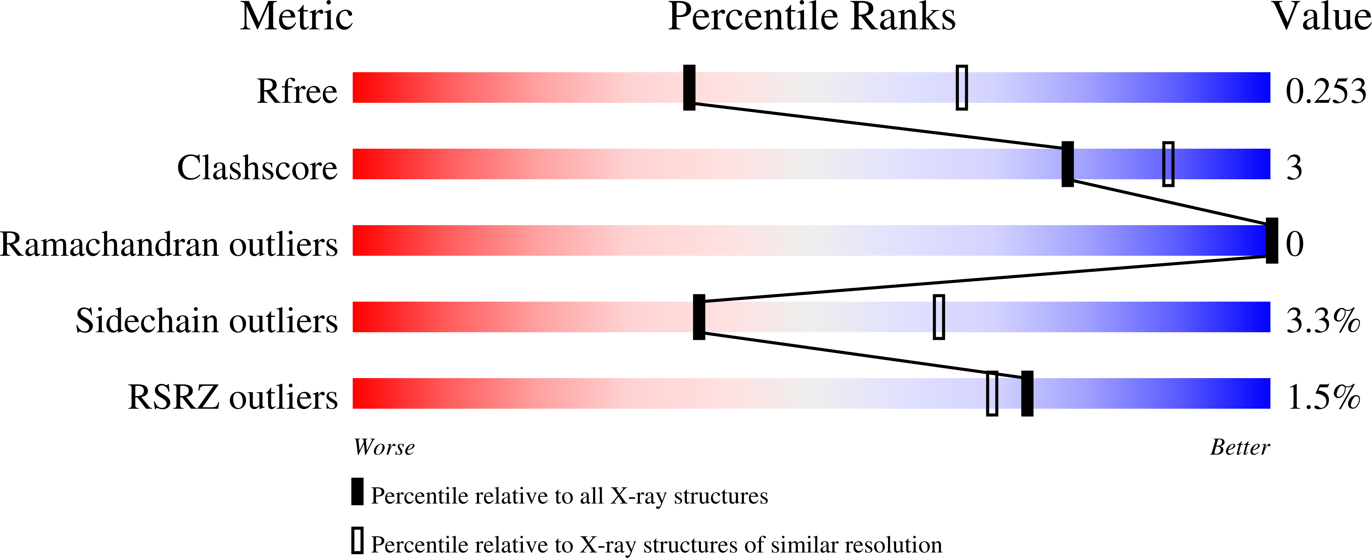
Experimental Data Snapshot
Starting Model: experimental
View more details
Entity ID: 1 | |||||
|---|---|---|---|---|---|
| Molecule | Chains | Sequence Length | Organism | Details | Image |
| Casein kinase II subunit alpha | 338 | Homo sapiens | Mutation(s): 0 Gene Names: CSNK2A1, CK2A1 EC: 2.7.11.1 | 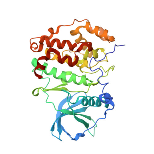 | |
UniProt & NIH Common Fund Data Resources | |||||
PHAROS: P68400 GTEx: ENSG00000101266 | |||||
Entity Groups | |||||
| Sequence Clusters | 30% Identity50% Identity70% Identity90% Identity95% Identity100% Identity | ||||
| UniProt Group | P68400 | ||||
Sequence AnnotationsExpand | |||||
Reference Sequence | |||||
| Ligands 2 Unique | |||||
|---|---|---|---|---|---|
| ID | Chains | Name / Formula / InChI Key | 2D Diagram | 3D Interactions | |
| X5E (Subject of Investigation/LOI) Download:Ideal Coordinates CCD File | H [auth A], M [auth B] | 4-[6-(6-propan-2-yloxyindol-1-yl)pyrazin-2-yl]benzoic acid C22 H19 N3 O3 FMENLVZROFNANR-UHFFFAOYSA-N |  | ||
| SO4 Download:Ideal Coordinates CCD File | C [auth A] D [auth A] E [auth A] F [auth A] G [auth A] | SULFATE ION O4 S QAOWNCQODCNURD-UHFFFAOYSA-L |  | ||
| Length ( Å ) | Angle ( ˚ ) |
|---|---|
| a = 127.842 | α = 90 |
| b = 127.842 | β = 90 |
| c = 125.341 | γ = 90 |
| Software Name | Purpose |
|---|---|
| REFMAC | refinement |
| Aimless | data scaling |
| XDS | data reduction |
| MOLREP | phasing |
| Funding Organization | Location | Grant Number |
|---|---|---|
| The Structural Genomics Consortium (SGC) | Canada | -- |


