8QMB | pdb_00008qmb

Nucleant-assisted 2.0 A resolution structure of the Streptococcus pneumoniae topoisomerase IV-V18mer DNA complex with the novel fluoroquinolone Delafloxacin


Experimental Data Snapshot

  • Method: X-RAY DIFFRACTION
  • Resolution: 2.00 Å
  • R-Value Free: 
    0.206 (Depositor), 0.184 (DCC) 
  • R-Value Work: 
    0.169 (Depositor) 

Starting Model: experimental
View more details

wwPDB Validation 3D Report Full Report

Validation slider image for 8QMB

Ligand Structure Quality Assessment 


This is version 1.1 of the entry. See complete history

Literature

Structural basis of topoisomerase targeting by delafloxacin.

Najmudin, S.Pan, X.S.Wang, B.Govada, L.Chayen, N.E.Rubio, N.Shaffer, M.S.P.Rzepa, H.S.Fisher, L.M.Sanderson, M.R.

(2025) Nat Commun 16: 5829-5829

  • DOI: https://doi.org/10.1038/s41467-025-60688-3
  • Primary Citation Related Structures: 
    8C41, 8QMB, 8QMC

  • PubMed Abstract: 

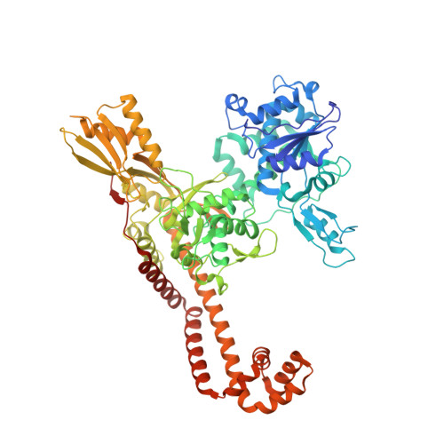
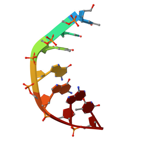
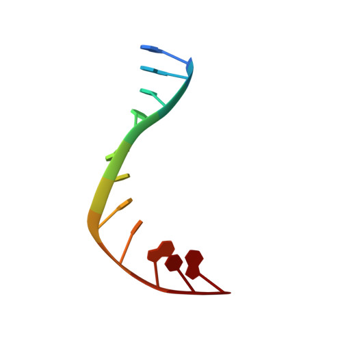
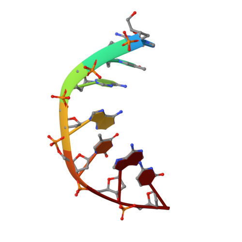
    Delafloxacin is a potent anionic fluoroquinolone approved for the treatment of respiratory infections that acts by trapping the DNA cleavage complexes of bacterial topoisomerase IV and gyrase. Its N-1-pyridinyl-, C-7-azetidinyl- and C-8-chlorine substituents confer enhanced antibiotic activity against bacteria resistant to other fluoroquinolones, but its mode of action is unclear. Here we present the X-ray crystal structures of a delafloxacin-DNA cleavage complex obtained by co-crystallization with Streptococcus pneumoniae topo IV using a graphene nucleant and solved at 2.0 and 2.4 Å resolution. The two Mg 2+ -chelated delafloxacin molecules intercalated at the DNA cleavage site are bound in an unusual conformation involving interacting out-of-plane N-1-aromatic- and C-8-chlorine- substituents. The unprecedented resolution allows comprehensive imaging of water-metal ion links integrating enzyme and DNA through drug-bound and active-site Mg 2+ ions plus the discovery of enzyme-bound K + ions. Our studies on delafloxacin action suggest that intrinsic target affinity contributes to its activity against quinolone-resistant bacteria.


  • Organizational Affiliation
    • Molecular and Cellular Sciences Section, Neuroscience and Cell Biology Research Institute, City St George's, University of London, Cranmer Terrace, London, UK.

Macromolecule Content 

  • Total Structure Weight: 181.44 kDa 
  • Atom Count: 13,475 
  • Modeled Residue Count: 1,484 
  • Deposited Residue Count: 1,520 
  • Unique protein chains: 1
  • Unique nucleic acid chains: 4

Macromolecules


Find similar proteins by:|  3D Structure
Entity ID: 1
MoleculeChains  Sequence LengthOrganismDetailsImage
DNA topoisomerase 4 subunit B,DNA topoisomerase 4 subunit A
A, B
742Streptococcus pneumoniaeMutation(s): 3 
Gene Names: parESP_0852parCSP_0855
EC: 5.6.2.2
UniProt
Find proteins for P72525 (Streptococcus pneumoniae serotype 4 (strain ATCC BAA-334 / TIGR4))
Explore P72525 
Go to UniProtKB:  P72525
Find proteins for Q59961 (Streptococcus pneumoniae serotype 4 (strain ATCC BAA-334 / TIGR4))
Explore Q59961 
Go to UniProtKB:  Q59961
Entity Groups
Sequence Clusters30% Identity50% Identity70% Identity90% Identity95% Identity100% Identity
UniProt GroupsQ59961P72525
Sequence Annotations
Expand
Reference Sequence
Find similar nucleic acids by:  Sequence
Entity ID: 2
MoleculeChains LengthOrganismImage
DNA (5'-D(TP*GP*TP*GP*GP*AP*T)-3')C [auth E]7Escherichia coli
Sequence Annotations
Expand
Reference Sequence
Find similar nucleic acids by:  Sequence
Entity ID: 3
MoleculeChains LengthOrganismImage
DNA (5'-D(P*GP*GP*TP*TP*AP*TP*CP*CP*AP*CP*A)-3')D [auth F]11Escherichia coli
Sequence Annotations
Expand
Reference Sequence
Find similar nucleic acids by:  Sequence
Entity ID: 4
MoleculeChains LengthOrganismImage
DNA (5'-D(GP*TP*AP*AP*TP*AP*C)-3')E [auth G]7Escherichia coli
Sequence Annotations
Expand
Reference Sequence
Find similar nucleic acids by:  Sequence
Entity ID: 5
MoleculeChains LengthOrganismImage
DNA (5'-D(P*AP*AP*CP*CP*GP*TP*AP*TP*TP*AP*C)-3')F [auth H]11Escherichia coli
Sequence Annotations
Expand
Reference Sequence

Small Molecules

Ligands 7 Unique
IDChains Name / Formula / InChI Key2D Diagram3D Interactions
TE9
(Subject of Investigation/LOI)

Query on TE9


Download:Ideal Coordinates CCD File
BA [auth F],
CA [auth H]
delafloxacin
C18 H12 Cl F3 N4 O4
DYDCPNMLZGFQTM-UHFFFAOYSA-N
MPD

Query on MPD


Download:Ideal Coordinates CCD File
K [auth A],
L [auth A],
Z [auth B]
(4S)-2-METHYL-2,4-PENTANEDIOL
C6 H14 O2
SVTBMSDMJJWYQN-YFKPBYRVSA-N
MLA

Query on MLA


Download:Ideal Coordinates CCD File
M [auth A],
O [auth A]
MALONIC ACID
C3 H4 O4
OFOBLEOULBTSOW-UHFFFAOYSA-N
ACT

Query on ACT


Download:Ideal Coordinates CCD File
N [auth A],
Q [auth A],
W [auth B],
X [auth B]
ACETATE ION
C2 H3 O2
QTBSBXVTEAMEQO-UHFFFAOYSA-M
FMT

Query on FMT


Download:Ideal Coordinates CCD File
AA [auth B],
P [auth A],
R [auth A],
Y [auth B]
FORMIC ACID
C H2 O2
BDAGIHXWWSANSR-UHFFFAOYSA-N
CL

Query on CL


Download:Ideal Coordinates CCD File
J [auth A],
V [auth B]
CHLORIDE ION
Cl
VEXZGXHMUGYJMC-UHFFFAOYSA-M
MG

Query on MG


Download:Ideal Coordinates CCD File
G [auth A]
H [auth A]
I [auth A]
S [auth B]
T [auth B]
G [auth A],
H [auth A],
I [auth A],
S [auth B],
T [auth B],
U [auth B]
MAGNESIUM ION
Mg
JLVVSXFLKOJNIY-UHFFFAOYSA-N

Experimental Data & Validation

Experimental Data

  • Method: X-RAY DIFFRACTION
  • Resolution: 2.00 Å
  • R-Value Free:  0.206 (Depositor), 0.184 (DCC) 
  • R-Value Work:  0.169 (Depositor) 
Space Group: P 31 2 1
Unit Cell:
Length ( Å )Angle ( ˚ )
a = 157.215α = 90
b = 157.215β = 90
c = 211.948γ = 120
Software Package:
Software NamePurpose
REFMACrefinement
DIALSdata reduction
xia2data scaling
STARANISOdata scaling
PHASERphasing

Structure Validation

View Full Validation Report


Ligand Structure Quality Assessment 


Entry History 

& Funding Information

Deposition Data


Funding OrganizationLocationGrant Number
Medical Research Council (MRC, United Kingdom)United KingdomMR/T000848/1

Revision History  (Full details and data files)

  • Version 1.0: 2024-10-02
    Type: Initial release
  • Version 1.1: 2025-07-23
    Changes: Database references, Structure summary